- 有料閲覧
- 文献概要
- 1ページ目
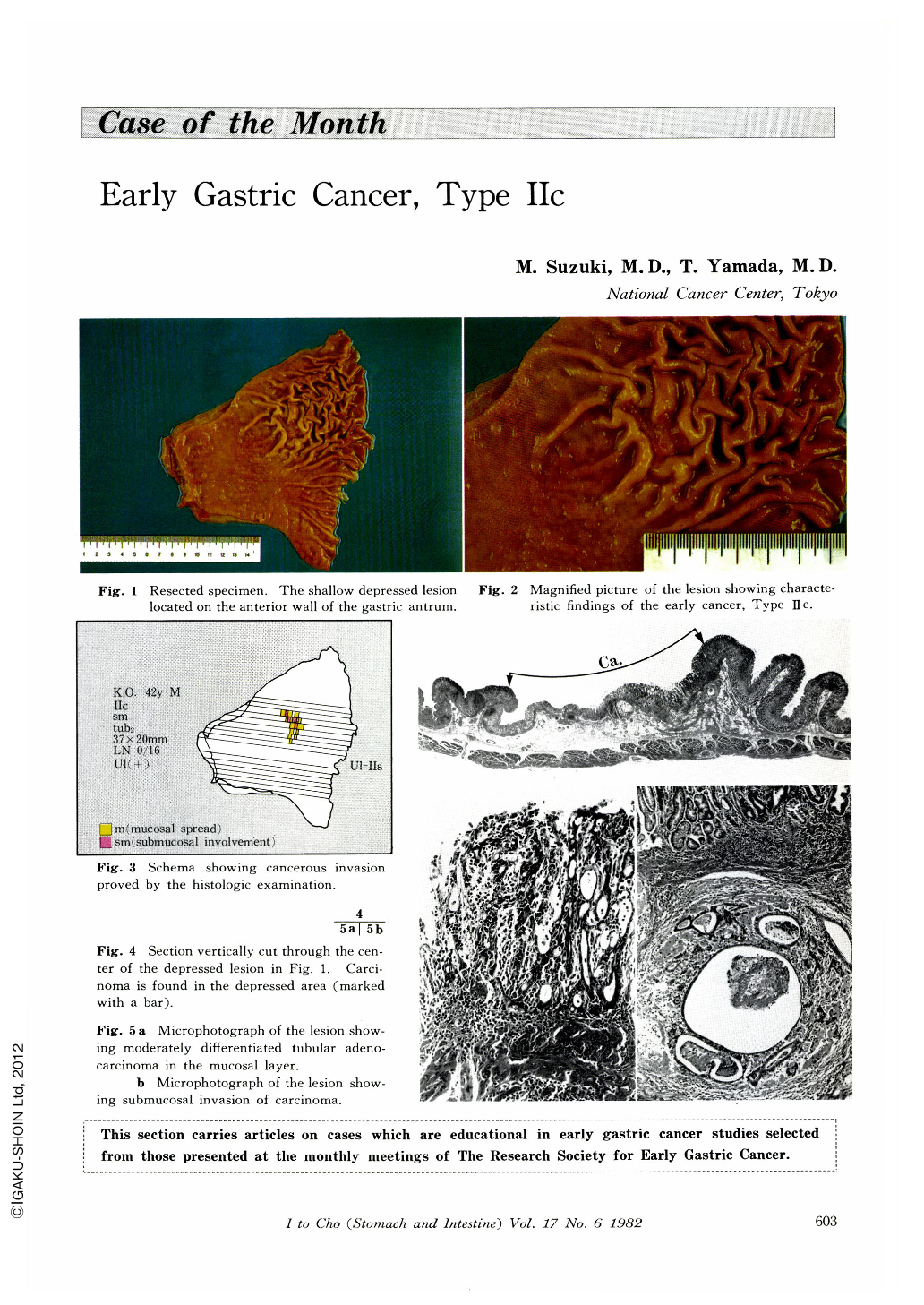
A 42-year-old man was admitted to the National Cancer Center Hospital for the detailed examination of the stomach because of abnormal findings detected in the gastric mass survery. Nothing notable was found in his personal and family history. Physical examination revealed no abnormal findings. Blood count, blood chemistry and urinalysis were within normal limits.
Radiography and endoscopy revealed an irregular, shallow depressed lesion with the converging folds on the anterior wall of the gastric antrum. Distinct tapering and the moth-eaten appearance was seen at the tips of the converging folds. But there was no such findings as clubbing and fusion of the folds, plateau-like elevation and irregularity of fine granular appearance within the depression, which are suggestive of submucosal involvement of cancer (sm).

Copyright © 1982, Igaku-Shoin Ltd. All rights reserved.


