Japanese
English
- 有料閲覧
- Abstract 文献概要
- 1ページ目 Look Inside
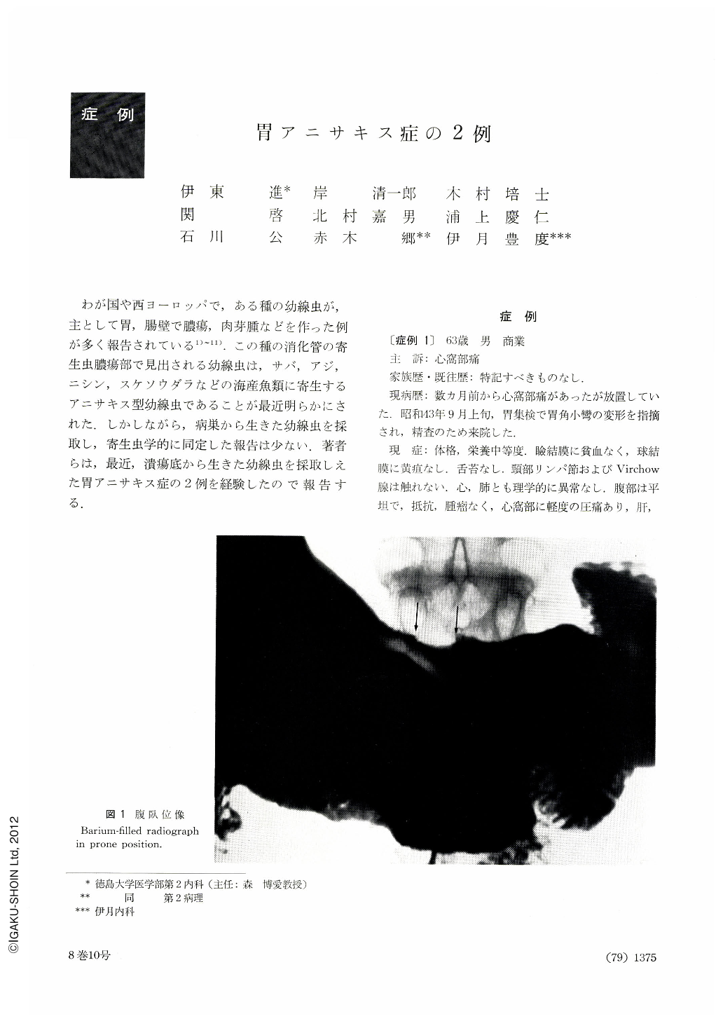
わが国や西ヨーロッパで,ある種の幼線虫が,主として胃,腸壁で膿瘍,肉芽腫などを作った例が多く報告されている1)~11).この種の消化管の寄生虫膿瘍部で見出される幼線虫は,サバ,アジ,ニシン,スケソウダラなどの海産魚類に寄生するアニサキス型幼線虫であることが最近明らかにされた.しかしながら,病巣から生きた幼線虫を採取し,寄生虫学的に同定した報告は少ない.著者らは,最近,潰瘍底から生きた幼線虫を採取しえた胃アニサキス症の2例を経験したので報告する.
This is a report of two recent cases of gastric anisakiasis in which the larvae were taken out of ulcer floor alive.
The first case: a 60-year-old merchant, suffering from episodes of epigastric pain since several months before. Gastric x-ray revealed a niche and irregularity of the gastric contour on the lesser curvature of the angle region. A shadow defect was also noticed on the pyloric side of the ulcer. Endoscopy further showed there a round ulcer with noticiable edema in the neighboring areas. On gastric resection, a still living anisakis larva was found on the bottom of the ulcer. On its distal side was also seen a protrusion suggestive of a submucosal tumor, later histologically confirmed as eosinophilic granuloma due to the parasite.
The second case: a male company employee aged 70 with bouts of epigastric pain for the past one month. X-ray of the stomach showed rugal convergency on the anterior wall side near the lesser curvature of the angle. An ulcer was revealed by endoscopy in its neighborhood. It diminished in size after one month, but an elevation likewise suggesting a submucosal tumor, hardly noticiable at the first examination, was distinctly observed in the side of the greater curvature. As in the first case, an anisakis larva was found on the ulcer floor. We managed to take it away out of the stomach with biopsy forceps under direct vision.
There have been quite a number of reports on tumor or granuloma mainly in the stomach or intestine brought about by a certain type of Nematoda, and now it has been clarified that anisakis larva parasitic on sea fish is responsible for the neoplasm. However, only a few concern with removal of a live larva out of a gastric lesion followed by parasitologic identification. In acute anisakiasis of the stomach, living larvae have been observed at times by endoscopy, but our case seems the first report on anisakiasis in which anisakis larva was taken out of the stomach under direct vision from a lesion such as abscess or granuloma. In this respect, our second report is of great interest to us.
Copyright © 1973, Igaku-Shoin Ltd. All rights reserved.