Japanese
English
- 有料閲覧
- Abstract 文献概要
- 1ページ目 Look Inside
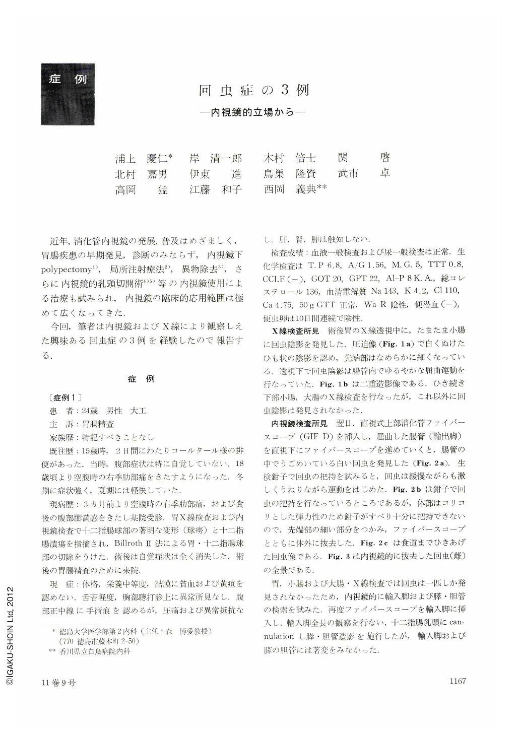
近年,消化管内視鏡の発展,普及はめざましく,胃腸疾患の早期発見,診断のみならず,内視鏡下polypectomy1),局所注射療法2),異物除去3),さらに内視鏡的乳頭切開術4)5)等の内視鏡使用による治療も試みられ,内視鏡の臨床的応用範囲は極めて広くなってきた.
今回,筆者は内視鏡およびX線により観察しえた興味ある回虫症の3例を経験したので報告する.
Owing to rapid progress in gastrointestinal endoscopy, it is increasingly used in clinical practice. It is, however, very rare to see ascaris during endoscopy, because ascariasis is seen only infrequently now in Japan. Three cases of ascariasis observed recently by endoscopy are considered worth reporting here.
Case 1. A 24-year-old male. During an examination of the upper G.I. series of the patient whose stomach had been resected by Billroth Ⅱ method, an ascaris was observed in the efferent loop about one m. distal from the stomach. Next day, by inserting GIF type D deep into the efferent loop, we found the worm again. Under fiberscopic observation it was held with biopsy forceps and taken out. Subsequent endoscopic observation followed by ERCP revealed no abnormality.
Case 2. A 40-year-old female. She was admitted to hospital because of severe upper abdominal pain with nausea and high fever. There was no jaundice. Five days later, the stomach was examined by JF-B, which showed no abnormality there. ERCP also revealed no abnormal changes in the pancreatic duct. In the bile duct, however, the shadow of an ascaris was observed. Her symptoms disappeared after ER CP. It was performed again two weeks later, but there was no shadow of ascaris this time.
Case 3. A 71-years-old female. She had complained of dull upper abdominal pain since a month before. X-ray study of the upper G.I. series revealed irregularity of the gastric angle. As following endoscopy of the stomach by JF-B revealed irregular depression of the angle, biopsy was performed. The instrument was then inserted further deep into the duodenum. ERCP showed no abnormality both in the bile and pancreatic ducts, but the shadow of an ascaris was de-monstrated by the contrast material (60% Urografin) overflowing to the jejunum. The histologic diagnosis of the biopsy specimens from the stomach was tubular adenocarcinoma. During the operation of the stomach one ml of tincture of iodine was injected into the body of the worm via the jejunal serosa. The ascaris was found in the stool one week later.

Copyright © 1976, Igaku-Shoin Ltd. All rights reserved.


