Japanese
English
- 有料閲覧
- Abstract 文献概要
- 1ページ目 Look Inside
症 例
患 者:竹○三○,64歳,男.
主 訴:自覚症状なし.
既往歴:特記すべきことなし.
家族歴:父親および弟が胃癌で死亡.
現病歴:昭和46年7月に札幌市がおこなった胃集検で異常を指摘され,当科で精密検査をおこない経過観察が必要であると説明された.健康には自信があったが約9カ月後の昭和47年3月,再度当科を訪れ精密検査の結果入院した.
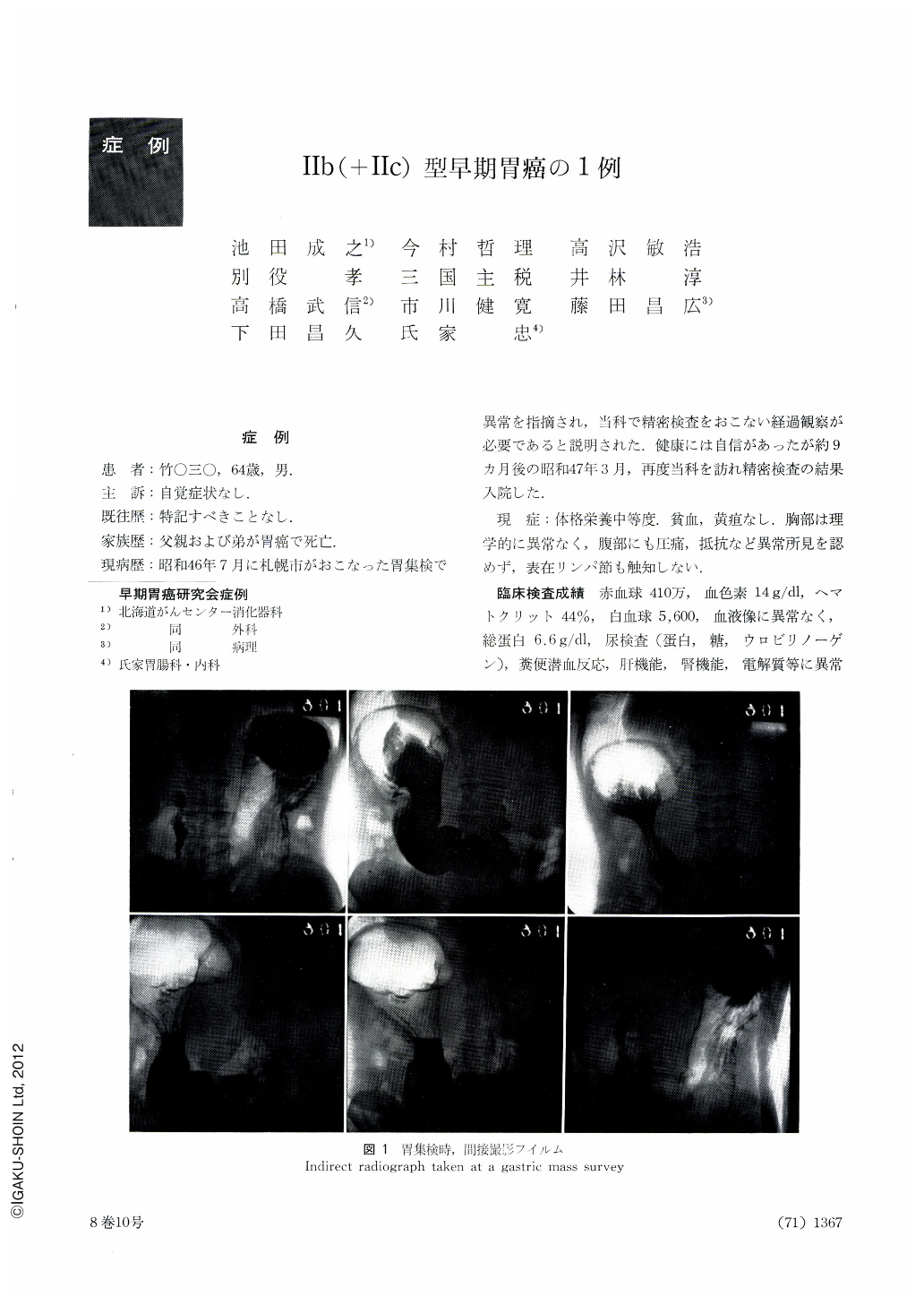
A 64-year-old man with no symptom to complain of was referred to our department because of an abnormality pointed out at a mass gastric scrreening. Indirect radiograms showed in barium-filled picture mural rigidity of the mid-body along with localized indentation, revealed in double contrast study, of the greater curvature of the same site. Opposite to it and along the lesser curvature was the gastric contour also rigid. Direct x-ray exposures in double contrast with small amount of air revealed a stricture on the lesser curvature of the midbody. Irregularity of the gastric contour was extensively seen from here down to the lower corpus. The areae gastricae over the area corresponding to that of constriction and irregular contour were flattened out. Barium adhered to this part in a way different from other portions.
Endoscopy revealed several small granular reddening on the lesser curvature side of the mid-body. The surface of the mucosa was slightly uneven and irregular. Nine months after the initial examination the patient had to undergo gastrectomy because carcinoma mucocellulare was revealed by biopsy in the mid-body. Gross observation of the resected stomach showed small granular reddening scattered about in the lesser curvature side of the mid-body, but there was not enough evidence, such as unevenness and difference in size of the areae gastricae, to warrant a suspicion of cancer. On closer scrutiny, however, one was barely able observe very slight depression in a part of the cancer lesion. Strictly speaking, it was a IIb-like finding. The areae disappeared after fixation. The mucosal surface looked more smooth as compared with other parts. The majority of the cancer nests showed a typical II b type, exhibiting no alteration on the mucosal surface as mentioned above. The entire lesion was diagnosed as IIb (+IIc).

Copyright © 1973, Igaku-Shoin Ltd. All rights reserved.


