Japanese
English
- 有料閲覧
- Abstract 文献概要
- 1ページ目 Look Inside
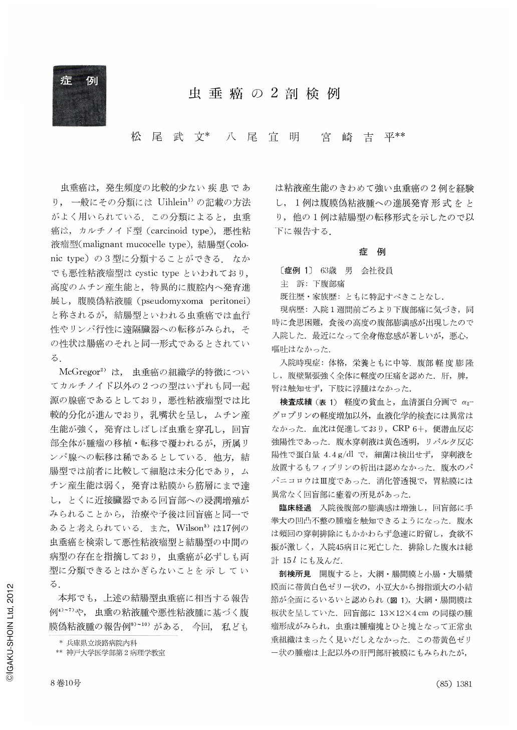
虫垂癌は,発生頻度の比較的少ない疾患であり,一般にその分類にはUihlein1)の記載の方法がよく用いられている.この分類によると,虫垂癌は,カルチノイド型(carcinoid type),悪性粘液瘤型(malignant mucocelle type),結腸型(colonic type)の3型に分類することができる.なかでも悪性粘液瘤型はcystic typeといわれており,高度のムチン産生能と,特異的に腹腔内へ発育進展し,腹膜偽粘液腫(pseudomyxoma peritonei)と称されるが,結腸型といわれる虫垂癌では血行性やリンパ行性に遠隔臓器への転移がみられ,その性状は腸癌のそれと同一形式であるとされている.
McGregor2)は,虫垂癌の組織学的特徴についてカルチノイド以外の2つの型はいずれも同一起源の腺癌であるとしており,悪性粘液瘤型では比較的分化が進んでおり,乳嘴状を呈し,ムチン産生能が強く,発育はしばしば虫垂を穿孔し,回盲部全体が腫瘤の移植・転移で覆われるが,所属リンパ腺への転移は稀であるとしている.他方,結腸型では前者に比較して細胞は未分化であり,ムチン産生能は弱く,発育は粘膜から筋層にまで達し,とくに近接臓器である回盲部への浸潤増殖がみられることから,治療や予後は回盲癌と同一であると考えられている.また,Wilson3)は17例の虫垂癌を検索して悪性粘液瘤型と結腸型の中間の病型の存在を指摘しており,虫垂癌が必ずしも両型に分類できるとはかぎらないことを示している.
Primary carcinoma of appendix is uncommon disease and divided into three types according to the pathological appearance; carcinoid type, malignant mucocelle type that produces pseudomyxoma peritonei and colonic type.
Two patients with pseudomucinous adenocarcinoma of appendical origin showing different mode in metastasis were reported. Case 1 is a typical case of malignant mucocelle which was complicated with pseudomyxoma peritonei, while case 2 is clinically a colonic type with massive liver metastasis but histologically is closer to case 1.
Case 1. A 63-year-old man was admitted to our clinic, complaining of lower abdominal pain of one week's duration. During hospitalization he underwent frequent abdominal paracentesis because of rapid accumulation of ascites. He deteriorated into cahexia and died. Postmortem examination revealed pseudomyxoma peritonei originating from appendix. Histologically it was ascertaind as adenocarcinoma with remarkable mucin production corresponding to malignant mucocelle type.
Case 2. A 68-year-old man was admitted to our clinic because of epigastric discomfort. Laboratory findings showed increased E. S. R., anemia and elevated serum alkaline phosphatase level. Postmortem examination showed cauliflower like tumor which probably originated from the mucosa of appendix and invaded into mucosa of cecum, and had large metastasis in the liver. Histologically adenocarcinoma containing much mucin was confirmed. Although histological appearance of case 2 was considered as malignant mucocelle type that resembled very much to that of case 1, it is interesting to note that the mode of metastasis was that of colonic type.

Copyright © 1973, Igaku-Shoin Ltd. All rights reserved.


