Japanese
English
- 有料閲覧
- Abstract 文献概要
- 1ページ目 Look Inside
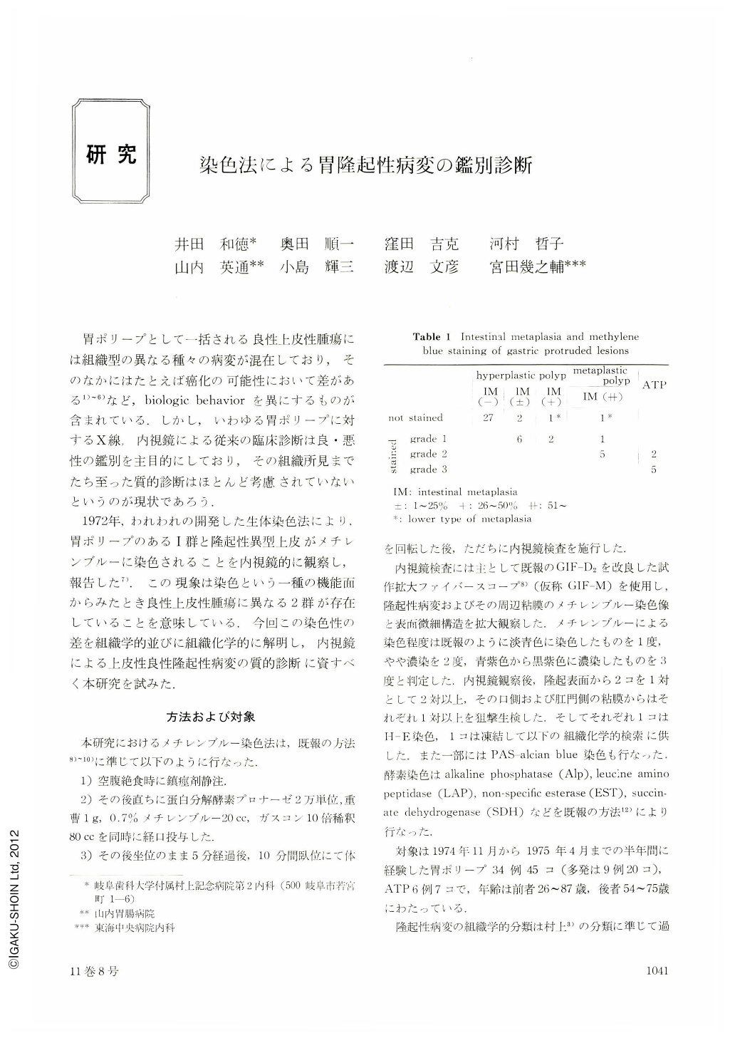
胃ポリープとして一括される良性上皮性腫瘍には組織型の異なる種々の病変が混在しており,そのなかにはたとえば癌化の可能性において差がある1)~6)など,biologic behaviorを異にするものが含まれている.しかし,いわゆる胃ポリープに対するX線,内視鏡による従来の臨床診断は良・悪性の鑑別を主目的にしており,その組織所見までたち至った質的診断はほとんど考慮されていないというのが現状であろう.
1972年,われわれの開発した生体染色法により,胃ポリープのあるⅠ群と隆起性異型上皮がメチレンブルーに染色されることを内視鏡的に観察し,報告した7).この現象は染色という一種の機能面からみたとき良性上皮性腫瘍に異なる2群が存在していることを意味している.今回この染色性の差を組織学的並びに組織化学的に解明し,内視鏡による上皮性良性隆起性病変の質的診断に資すべく本研究を試みた.
In 1972, we reported that a certain group of gastric polyp and atypical epithelium was stained with methylene blue in vivo using our staining method. In this paper we report morphological, histological and histochemical characteristics of these stained lesions.
1) Among 45 polyps of 34 cases, 14 polyps of 12 cases were stained with methylene blue. These stained polyps consisted of hyperplastic polyps with metaplasia and polyps of intestinal epithelium. The more remarkable intestinal metaplasia accompanied polyps, the darker they were stained. All benign atypical epitheliums were stained dark blue. Namely, polyp with intestinal metapasia and atypical epithelium which has a malignant potential can be differentiated from non-metaplastic, hyperplastic polyp with endoscopy.
2) There was no difference in histochemistory between the intestinal metaplasias of polyp and gastric mucosa. Therefore, it seems likely that the staining of gastric polyp is based on absorption of methylene blue in the same manner as in intestinalized gastric mucosa.
3) With magnifying fiberscope, it was observed that the surface of stained polyp was covered with the mucosa resembling intestinal metaplasia of gastric mucosa.
4) As the average age of patients with polyp advanced, its metaplasia became more remarkable.
5) Although non-metaplastic, hyperplastic polyps were found in any part of the stomach, most of metaplastic, hyperplastic polyps and polyps of intestinal epithelium were found in the antrum covered with intestinalized mucosa.

Copyright © 1976, Igaku-Shoin Ltd. All rights reserved.


