Japanese
English
- 有料閲覧
PPV購入案内
1,320円 (1,200円+税10%) こちらは、941ページから944ページの文献です。購入には医書.jp本会員登録が必要です。
会員登録完了後に改めて上記「購入サイトへ」を選択してください。
または「購入サイトへ」選択後に「購入ログイン」、「新規会員登録」の順に進んでください。
会員登録について詳しくはをご確認ください。
- Abstract 文献概要
- 1ページ目 Look Inside
- 参考文献 Reference
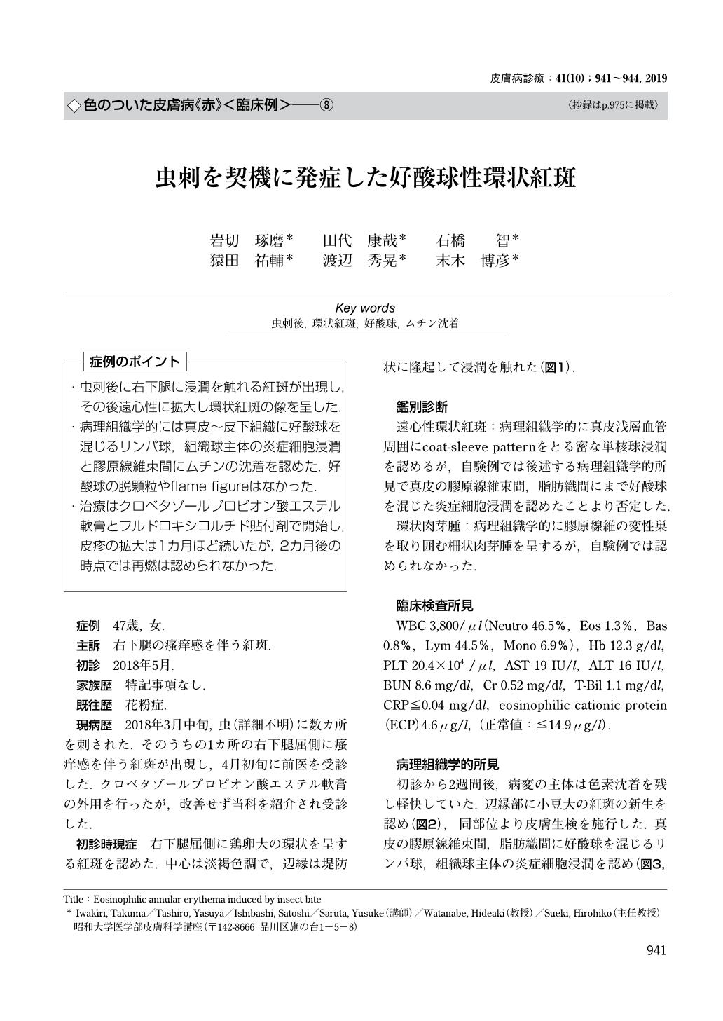
・虫刺後に右下腿に浸潤を触れる紅斑が出現し,その後遠心性に拡大し環状紅斑の像を呈した.
・病理組織学的には真皮〜皮下組織に好酸球を混じるリンパ球,組織球主体の炎症細胞浸潤と膠原線維束間にムチンの沈着を認めた.好酸球の脱顆粒やflame figureはなかった.
・治療はクロベタゾールプロピオン酸エステル軟膏とフルドロキシコルチド貼付剤で開始し,皮疹の拡大は1カ月ほど続いたが,2カ月後の時点では再燃は認められなかった.
(「症例のポイント」より)
Eosinophilic annular erythema induced-by insect bite
Iwakiri, Takuma1)Tashiro, Yasuya1)Ishibashi, Satoshi1)Saruta, Yusuke1)Watanabe, Hideaki1)Sueki, Hirohiko1) 1)Department of Dermatology, Showa University School of Medicine
A 47-year-old woman visited dermatology department complaining of pruritic erythema at her right leg. In mid-March 2018, she was bitten by an insect. An erythematous plaque appeared on the same site, and then expanded centrifugally. Examination revealed erythematous plaque with a hen-egg-sized ring was observed on the flexor surface of the right lower leg. The center was light brown, and the rim was raised like a dike. Biopsy revealed lymphocytic infiltration, admixing eosinophils and histiocytes between the collagen bundles of the dermis and subcutaneous tissue. Mucin deposition was observed between the collagen bundles. Eosinophil degranulation and flame figures were not observed. High potent steroid was applied under the final diagnosis of eosinophilic annular erythema. The skin lesion continued for about a month, but no relapse was observed after 2 months.
Copyright © 2019, KYOWA KIKAKU Ltd. All rights reserved.