Japanese
English
- 有料閲覧
PPV購入案内
1,320円 (1,200円+税10%) こちらは、1283ページから1287ページの文献です。購入には医書.jp本会員登録が必要です。
会員登録完了後に改めて上記「購入サイトへ」を選択してください。
または「購入サイトへ」選択後に「購入ログイン」、「新規会員登録」の順に進んでください。
会員登録について詳しくはをご確認ください。
- Abstract 文献概要
- 1ページ目 Look Inside
- 参考文献 Reference
はじめに
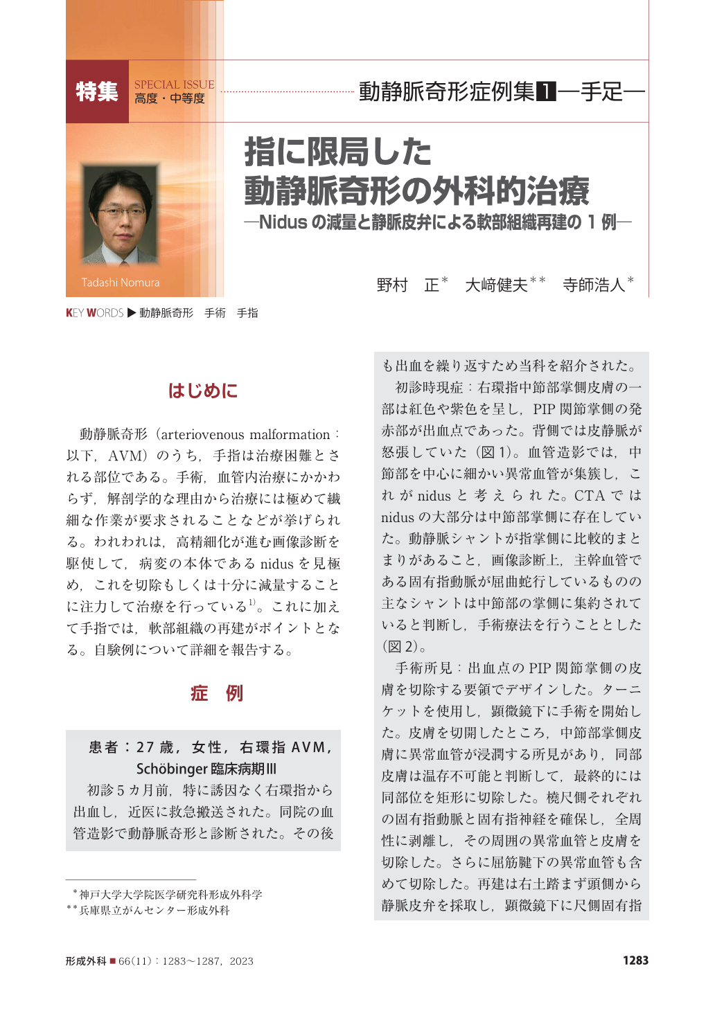
動静脈奇形(arteriovenous malformation:以下,AVM)のうち,手指は治療困難とされる部位である。手術,血管内治療にかかわらず,解剖学的な理由から治療には極めて繊細な作業が要求されることなどが挙げられる。われわれは,高精細化が進む画像診断を駆使して,病変の本体であるnidusを見極め,これを切除もしくは十分に減量することに注力して治療を行っている 1)。これに加えて手指では,軟部組織の再建がポイントとなる。自験例について詳細を報告する。
Among the sites at which arteriovenous malformations (AVMs) have been documented, an AVM in a finger is difficult to cure. Whether surgery or endovascular treatment is administered, extremely delicate work is required for treatment in light of the anatomical complexity of the fingers. We make full use of high-resolution imaging to identify the nidus, i.e., the main body of an AVM lesion, and we focus on resecting it or sufficiently reducing its volume. The reconstruction of the soft tissue is also a key point for fingers. We describe the case of a 27-year-old woman with a localized AVM in the right ring finger who underwent a microscopic excision and reconstruction with a venous flap. Five years after the operation, there is no obvious re-expansion. Surgery for an AVM can preserve a finger if the lesion is limited to the finger; such surgery can sufficiently reduce the nidus with proper soft-tissue reconstruction.
Copyright© 2023 KOKUSEIDO CO., LTD. All Rights Reserved.