Japanese
English
症例
EOB-MRIの肝細胞相で腫瘤周囲に広範な低信号域を呈した肝内胆管癌の1例
A case of intrahepatic cholangiocarcinoma showing broad peritumoral hypointensity on hepatobiliary phase in EOB-MRI
熊澤 秀亮
1
,
伊藤 茂樹
1
,
加賀谷 理紗
1
,
河合 雄一
1
,
館 靖
1
,
森 雄司
1
,
荒川 拓也
2
,
髙田 莉央
3
Shusuke Kumazawa
1
1日本赤十字社愛知医療センター名古屋第一病院 放射線診断科
2同 外科
3同 病理部
1Department of Diagnostic Radiology Japanese Red Cross Aichi Medical Center Nagoya Daiichi Hospital
キーワード:
肝内胆管癌
,
EOB-MRI
,
微小門脈侵襲
Keyword:
肝内胆管癌
,
EOB-MRI
,
微小門脈侵襲
pp.441-446
発行日 2023年5月10日
Published Date 2023/5/10
DOI https://doi.org/10.18888/rp.0000002325
- 有料閲覧
PPV購入案内
1,650円 (1,500円+税10%) こちらは、441ページから446ページの文献です。
PPV(ペイ・パー・ビュー)とは、論文(記事)単位で購入・閲覧ができるサービスです。
購入には医書.jp本会員登録が必要です。
会員登録完了後に改めて上記「購入サイトへ」を選択してください。
または「購入サイトへ」選択後に「購入ログイン」、「新規会員登録」の順に進んでください。
会員登録について詳しくはをご確認ください。
購入には医書.jp本会員登録が必要です。
会員登録完了後に改めて上記「購入サイトへ」を選択してください。
または「購入サイトへ」選択後に「購入ログイン」、「新規会員登録」の順に進んでください。
会員登録について詳しくはをご確認ください。
- Abstract 文献概要
- 1ページ目 Look Inside
- 参考文献 Reference
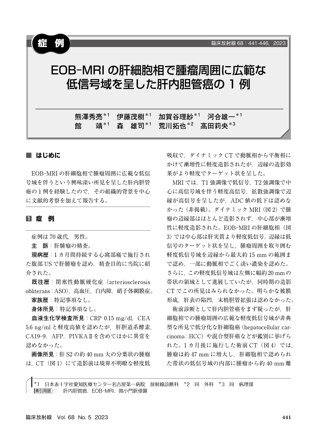
EOB-MRIの肝細胞相で腫瘤周囲に広範な低信号域を伴うという興味深い所見を呈した肝内胆管癌の1例を経験したので,その組織的背景を中心に文献的考察を加えて報告する。
The patient is a 70’s-year-old man. The liver mass was hypovascular at the margins and progressively mildly enhanced in the center on dynamic CT/MRI, and EOB-MRI showed broad peritumoral hypointensity on the hepatobiliary phase. There was also band-shaped hypointensity developing from the mass, within which a metastasis appeared preoperatively. The histopathological diagnosis was poorly differentiated intrahepatic cholangiocarcinoma with severe microportal invasion and secondary portal vein dilatation around the mass. This image finding can be useful for predicting microportal invasion in intrahepatic cholangiocarcinoma.
Copyright © 2023, KANEHARA SHUPPAN Co.LTD. All rights reserved.