Japanese
English
- 有料閲覧
PPV購入案内
1,320円 (1,200円+税10%) こちらは、731ページから740ページの文献です。購入には医書.jp本会員登録が必要です。
会員登録完了後に改めて上記「購入サイトへ」を選択してください。
または「購入サイトへ」選択後に「購入ログイン」、「新規会員登録」の順に進んでください。
会員登録について詳しくはをご確認ください。
- Abstract 文献概要
- 1ページ目 Look Inside
- 参考文献 Reference
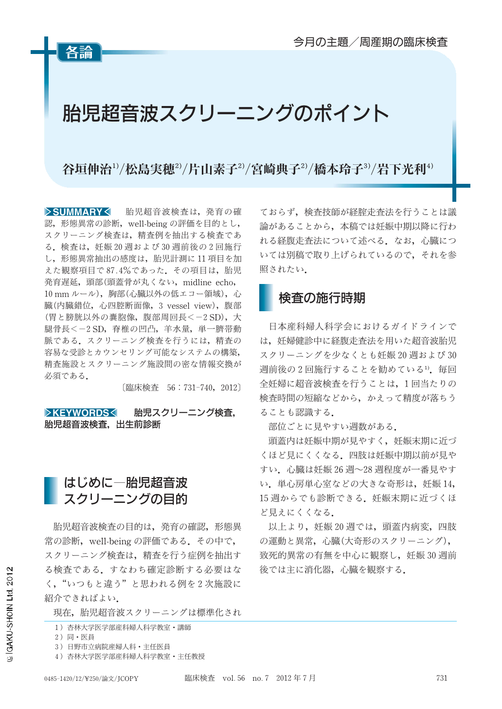
胎児超音波検査は,発育の確認,形態異常の診断,well-beingの評価を目的とし,スクリーニング検査は,精査例を抽出する検査である.検査は,妊娠20週および30週前後の2回施行し,形態異常抽出の感度は,胎児計測に11項目を加えた観察項目で87.4%であった.その項目は,胎児発育遅延,頭部(頭蓋骨が丸くない,midline echo,10mmルール),胸部(心臓以外の低エコー領域),心臓(内臓錯位,心四腔断面像,3 vessel view),腹部(胃と膀胱以外の囊胞像,腹部周回長<-2SD),大腿骨長<-2SD,脊椎の凹凸,羊水量,単一臍帯動脈である.スクリーニング検査を行うには,精査の容易な受診とカウンセリング可能なシステムの構築,精査施設とスクリーニング施設間の密な情報交換が必須である.
Ultrasound examination of the fetus attempts to assessment of fetal growth, identify structural anomalies, and evaluation of well-being. Fetal ultrasound screening test is carried out at 20 and 30 weeks gestation in order to find out the case which thorough examination. Eleven fetal ultrasound views which added fetal biometry are acquired in the fetal ultrasound screening test and sensitivity was 87.4%. In order to conduct screening test, the system in which easy consultation, counseling of thorough examination and dense information exchange between a thorough examination institution and a screening test institution is required.
Copyright © 2012, Igaku-Shoin Ltd. All rights reserved.