Japanese
English
- 有料閲覧
PPV購入案内
1,320円 (1,200円+税10%) こちらは、93ページから98ページの文献です。購入には医書.jp本会員登録が必要です。
会員登録完了後に改めて上記「購入サイトへ」を選択してください。
または「購入サイトへ」選択後に「購入ログイン」、「新規会員登録」の順に進んでください。
会員登録について詳しくはをご確認ください。
- Abstract 文献概要
- 1ページ目 Look Inside
I.緒言
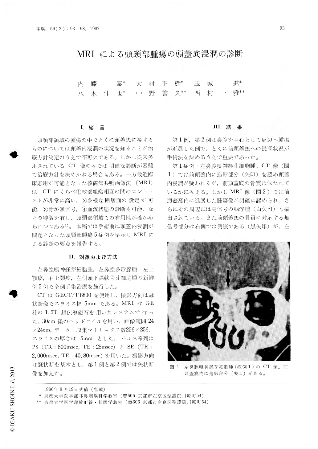
頭頸部領域の腫瘍の中でとくに頭蓋底に接するものについては頭蓋内浸潤の状況を知ることが治療方針決定のうえで不可欠である。しかし従来多用されているCT像のみでは明確な診断が困難で治療方針を決めかねる場合もある。一方最近臨床応用が可能となった核磁気共鳴画像法(MRI)は,CTにくらべ①軟部組織相互の間のコントラストが非常に高い,②多様な断層面の設定が可能,③骨が無信号,④血流状態の診断も可能,などの特徴を有し,頭頸部領域での有用性が確かめられつつある1)。本稿では手術前に頭蓋内浸潤が問題となった頭頸部腫瘍5症例を呈示しMRIによる診断の要点を報告する。
Magnetic resonance (MR) studies were performed on five patients of head and neck tumor with or without skull base invasions. In MRI, discontinuity of no signal line of skull base compact bone and a loss of high signal intensity of the fatty bone marrow indicate skull base destruction. Direct corona] and sagittal views can be obtained readily in MRI, and these are of great help to know the exact tumor extent. MRI is useful for the evaluation of skull base tumor invasion and for planning of its treatment.
Copyright © 1987, Igaku-Shoin Ltd. All rights reserved.