Japanese
English
- 有料閲覧
PPV購入案内
1,320円 (1,200円+税10%) こちらは、45ページから49ページの文献です。購入には医書.jp本会員登録が必要です。
会員登録完了後に改めて上記「購入サイトへ」を選択してください。
または「購入サイトへ」選択後に「購入ログイン」、「新規会員登録」の順に進んでください。
会員登録について詳しくはをご確認ください。
- Abstract 文献概要
- 1ページ目 Look Inside
I.はじめに
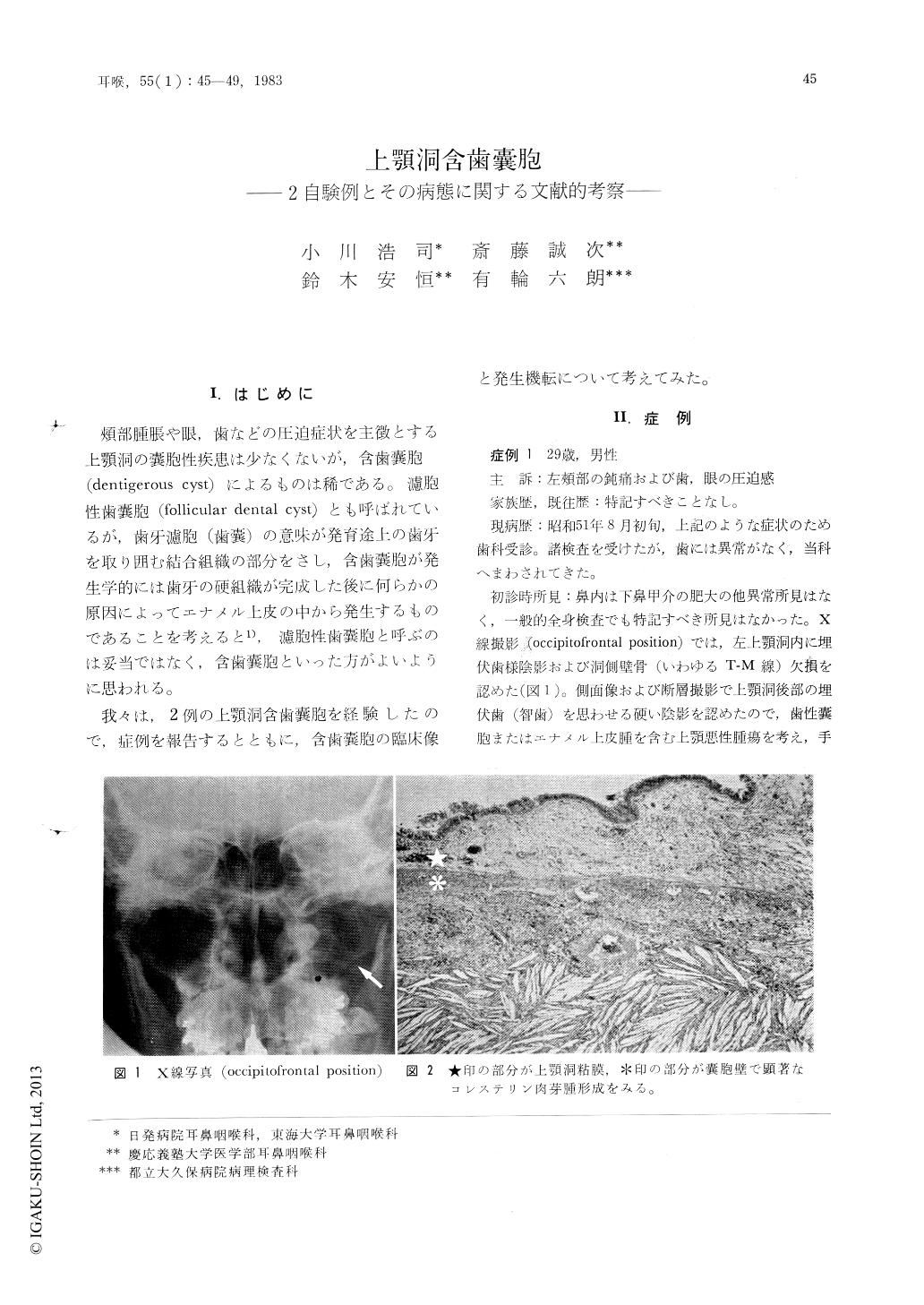
頬部腫脹や眼,歯などの圧迫症状を主徴とする上顎洞の嚢胞性疾患は少なくないが,含歯嚢胞(dentigerous cyst)によるものば稀である。濾胞性歯嚢胞(fbllicular dental cyst)とも呼ばれているが,歯牙濾胞(歯嚢)の意味が発育途上の歯牙を取り囲む結合組織の部分をさし,含歯嚢胞が発生学的には歯牙の硬組織が完成した後に何らかの原因によってエナメル上皮の中から発生するものであることを考えると1),濾胞性歯嚢胞と呼ぶのは妥当ではなく,含歯嚢胞といった方がよいように思われる。
我々は,2例の上顎洞含歯嚢胞を経験したので,症例を報告するとともに,含歯嚢胞の臨床像と発生機転について考えてみた。
Two cases of large dentigerous cyst involving the maxillary sinus were reported and discussion about clinical features and pathogenesis was also made. The first, a 29-year-old man, showed the half-erupted third molar within the maxillary sinus. The posterior lateral bony wall of the sinus was destroyed by pressure of the growing cyst. The cyst of the second case, an 8-year-old boy, was associated with an impacted canine tooth. The original small maxillary sinus with thin bony wall located behind the cyst. Histopathological examinations showed both of them to be dentigerous cyst. Dentigerous cyst appeared to be a sequel to impeded eruption or inflammation of the surrounding tissue.
Copyright © 1983, Igaku-Shoin Ltd. All rights reserved.