Japanese
English
- 有料閲覧
PPV購入案内
1,320円 (1,200円+税10%) こちらは、511ページから516ページの文献です。購入には医書.jp本会員登録が必要です。
会員登録完了後に改めて上記「購入サイトへ」を選択してください。
または「購入サイトへ」選択後に「購入ログイン」、「新規会員登録」の順に進んでください。
会員登録について詳しくはをご確認ください。
- Abstract 文献概要
- 1ページ目 Look Inside
I.はじめに
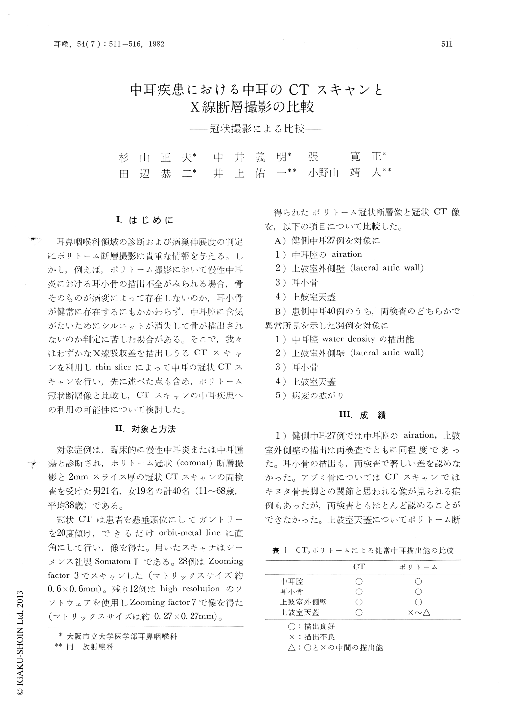
耳鼻咽喉科領域の診断および病巣伸展度の判定にポリトーム断層撮影は貴重な情報を与える。しかし,例えば,ポリトーム撮影において慢性中耳炎における耳小骨の描出不全がみられる場合,骨そのものが病変によって存在しないのか,耳小骨が健常に存在するにもかかわらず,中耳腔に含気がないためにシルエットが消失して骨が描出されないのか判定に苦しむ場合がある。そこで,我々はわずかなX線吸収差を描出しうるCTスキャンを利用しthin sliceによって中耳の冠状CTスキャンを行い,先に述べた点も含め,ポリトーム冠状断層像と比較し,CTスキャンの中耳疾患への利用の可能性について検討した。
We retrospectively analysed the coronal images of the middle ear obtained by multidirectional tomography (polytomography) and computed tomography (CT) in 40 patients. Although CT was capable of demonstrating water density in the middle ear more clearly than polytomography and of delineating a lesion extending even outside of the petrous bone, the diagnostic capability was not much different between the two tomographic techniques. On the other hand, coronal CT scan has a disadvantage in that it usually has to be performed during hyperextension of the neck or while patients are in an uncomfortable hanging head position. We think that CT scan should be utilized only in case with a lesion extending beyond the petrous bone and/or is not well visualized by polytomography.
Copyright © 1982, Igaku-Shoin Ltd. All rights reserved.