Japanese
English
- 有料閲覧
- Abstract 文献概要
- 1ページ目 Look Inside
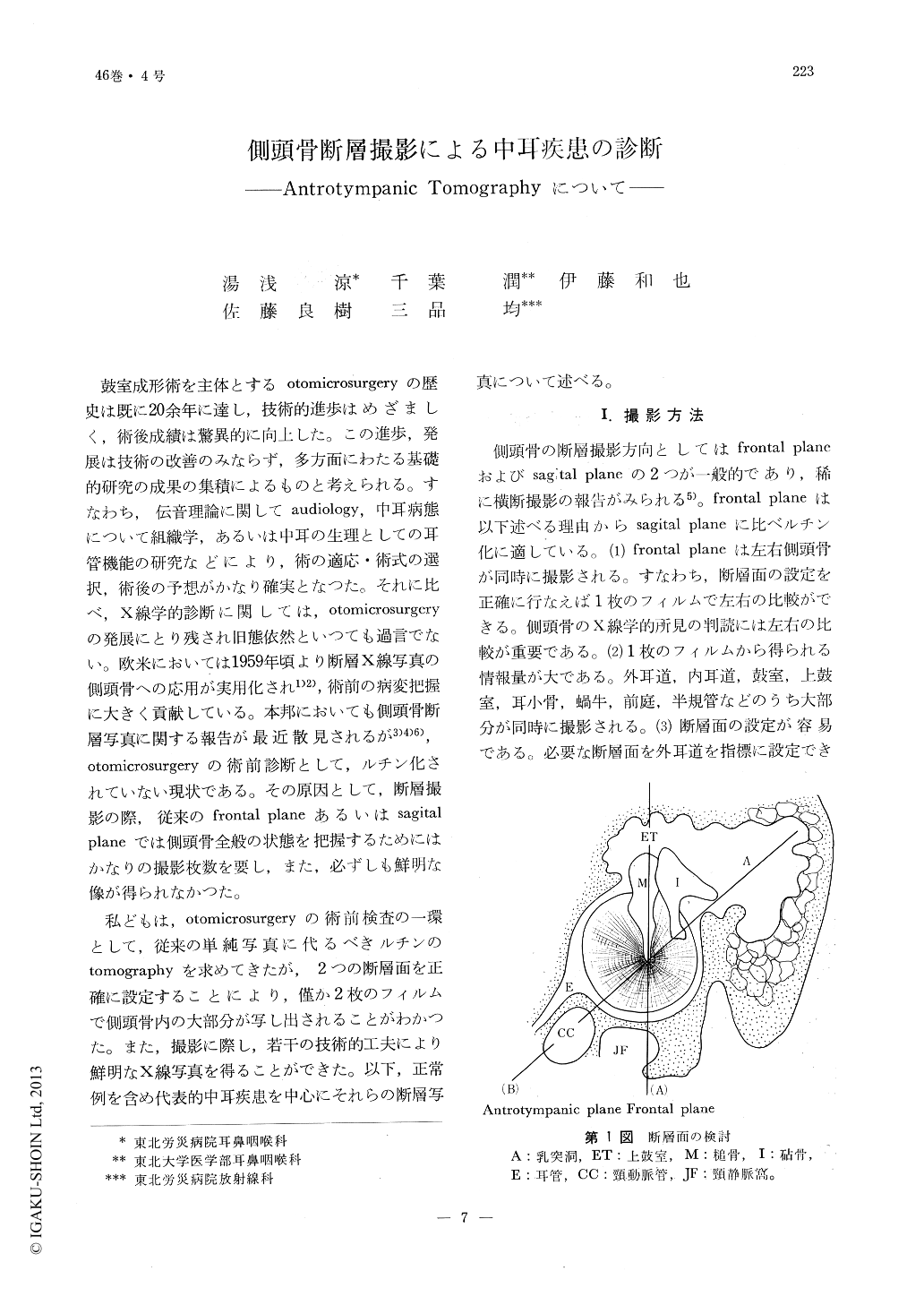
鼓室成形術を主体とするotomicrosurgeryの歴史は既に20余年に達し,技術的進歩はめざましく,術後成績は驚異的に向上した。この進歩,発展は技術の改善のみならず,多方面にわたる基礎的研究の成果の集積によるものと考えられる。すなわち,伝音理論に関してaudiology,中耳病態について組織学,あるいは中耳の生理としての耳管機能の研究などにより,術の適応・術式の選択,術後の予想がかなり確実となつた。それに比べ,X線学的診断に関しては,otomicrosurgeryの発展にとり残され旧態依然といつても過言でない。欧米においては1959年頃より断層X線写真の側頭骨への応用が実用化され1)2),術前の病変把握に大きく貢献している。本邦においても側頭骨断層写真に関する報告が最近散見されるが3)4)6),otomicrosurgeryの術前診断として,ルチン化されていない現状である。その原因として,断層撮影の際,従来のfrontal planeあるいはsagital planeでは側頭骨全般の状態を把握するためにはかなりの撮影枚数を要し,また,必ずしも鮮明な像が得られなかつた。
私どもは,otomicrosurgeryの術前検査の一環として,従来の単純写真に代るべきルチンのtomographyを求めてきたが,2つの断層面を正確に設定することにより,僅か2枚のフィルムで側頭骨内の大部分が写し出されることがわかつた。また,撮影に際し,若干の技術的工夫により鮮明なX線写真を得ることができた。以下,正常例を含め代表的中耳疾患を中心にそれらの断層写真について述べる。
With the advent of roulette tomography the x-ray diagnosis in the field of ENT has been greatly enhanced. But, due to the large number of plates that this procedure require it has notbeen popularized.
The authors succeeded in reducing the number of x-ray plates to that of two by first making the usual frontal plane exposure which is followed by another with antrotympanic plane. The latter is accomplished by turning the plane of exposure 35 degrees posteriorly. By these two plates all structures related to the middle ear as well as the carotid canal and the jugular foramen are clearly discernible.

Copyright © 1974, Igaku-Shoin Ltd. All rights reserved.


