Japanese
English
- 有料閲覧
- Abstract 文献概要
- 1ページ目 Look Inside
I.はじめに
中耳炎の診断と治療に際して,各症例の病態を詳細に把握するためには,病歴・耳鏡所見・各種聴力検査・X線所見などを総合的に判断する。慢性中耳炎に対する手術においては各種病態の処理法や手術手技などが進歩し種々の方法が採用されており,各症例の術前の詳細な評価はより有効でかつ必要なものとなつてきている。
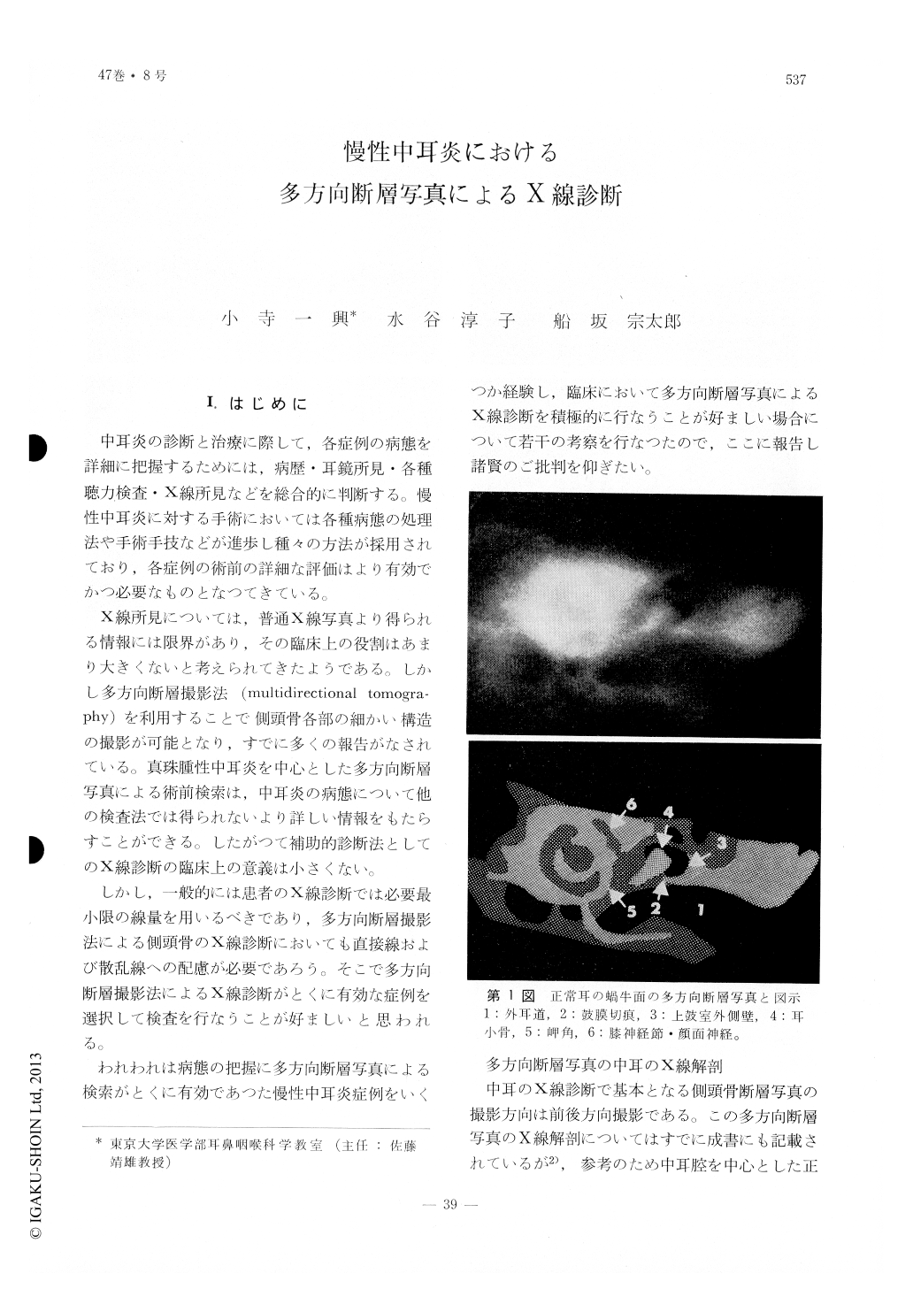
X線所見については,普通X線写真より得られる情報には限界があり,その臨床上の役割はあまり大きくないと考えられてきたようである。しかし多方向断層撮影法(multidirectional tomography)を利用することで側頭骨各部の細かい構造の撮影が可能となり,すでに多くの報告がなされている。真珠腫性中耳炎を中心とした多方向断層写真による術前検索は,中耳炎の病態について他の検査法では得られないより詳しい情報をもたらすことができる。したがつて補助的診断法としてのX線診断の臨床上の意義は小さくない。
Multidirectional tomographic studies of the temporal bone were made on 4 cases of chronic otitis media.
The first case was affected with chronic otitis media with a central perforation. Cholesteatoma was found by tomographic examination.
In the second case the cholesteatoma was found to be limited to the epitympnum which was verified later at the operation.
The third case presented a facial paralysis. Tomography revealed bone destruction at the geniculate ganglion.
Tomographic examination in the fourth case revealed the loss of malleus and incus.
The authors emphasize that multidirectional tomography is a means which is indispensable in evaluating cases of chronic otitis media.

Copyright © 1975, Igaku-Shoin Ltd. All rights reserved.


