Japanese
English
- 有料閲覧
PPV購入案内
1,320円 (1,200円+税10%) こちらは、139ページから144ページの文献です。購入には医書.jp本会員登録が必要です。
会員登録完了後に改めて上記「購入サイトへ」を選択してください。
または「購入サイトへ」選択後に「購入ログイン」、「新規会員登録」の順に進んでください。
会員登録について詳しくはをご確認ください。
- Abstract 文献概要
- 1ページ目 Look Inside
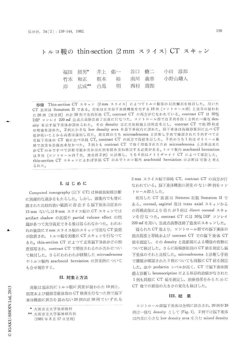
抄録 Thin-section-CTスキャン(2mmスライス)によつてトルコ鞍部の局所解剖を検討した。用いたCT装置はSomatom IIである。対象は正常脳下垂体機能を有する19例(コントロール群)と異常の疑われた20例(異常群)の計39例で全例単純CT,contrast CTの両方が行なわれている。contrast CTは30%DIPコンレイ220ml急速点滴静注終了直後に行なつた。コントロール群では2例を除く全例に一様なden—sityを示す脳下垂休が認められた。そのdensityは正常脳組織と同程度を示し,contrast CTで約25程度の増強を認めた。2例に小さなlow density areaを脳下垂体内に認めた。脳下垂体は海綿静脈洞に比べCT値が低いことから両者は識別し得た。異常群のうちmicroadenomaと診断し手術で確認された3例すべて正常脳下垂体のCT値に比べ単純CT,contrast CTの両方で高値を示した。3例のうち1例はポリトーム断層で異常を指摘出来なかつた。3例ともcontrast CTで強く増強されたためmicroadenomaと診断出来たがCTのみですべて診断可能か否かは症例を積み重ね検討する必要がある。トルコ鞍内arachnoid herniationは9例(コントロール群7例,異常群2例)に診断し,うち4例はメトリザマイドCTによつて確証した。thin-section CTスキャンによれば単純CTのみでトルコ鞍内arachnoid herniationの診断は可能と考えられた。
Topographic anatomy of the pituitary fossa was studied by 2mm thin-section CT scan (Somatom II). Nineteen with normal pituitary (control group) and 20 with suspected pituitary abnormality were se-lected. Plain and contrast CT were performed in all cases. Contrats CT was carried out immediately after the rapid infusion of 220ml of 30% iodinated contrast medium.
In all of control group but two, pituitary gland was detected as homogenous density and its density was as same as density of normal brain tissues, and was enhanced in degree of about 25CT number. In 2 cases, small low density was visualized in the pituitary gland. Pituitary gland was differentiated from cavernous sinus was usually higher than thepituitary gland.
In abnormal group, the microadenoma of the pituitary gland was diagnosed in 5 cases and 3 out of 5 cases was proved by surgery. All 3 micro-adenomas proved slightly dense by plain CT and enhanced higher than normal pituitary gland by contrast CT. Polytomograms showed no abnormal-ity of the sella turcica in one of these 3 cases. Although 3 microadenomas were detected by the abnormal enhancement, we are not sure whether all of microadenoma can be detected by CT alone.
Arachnoid herniation into the pituitary fossa was diagnosed in 7 of control group and 2 of abnormal group. Four out of these 9 cases were verified by using Metrizamide CT. By plain thin-section CT, the diagnosis of arachnoid herniation seems to be possible without Metrizamide CT.
Copyright © 1982, Igaku-Shoin Ltd. All rights reserved.