Japanese
English
- 有料閲覧
PPV購入案内
1,320円 (1,200円+税10%) こちらは、403ページから407ページの文献です。購入には医書.jp本会員登録が必要です。
会員登録完了後に改めて上記「購入サイトへ」を選択してください。
または「購入サイトへ」選択後に「購入ログイン」、「新規会員登録」の順に進んでください。
会員登録について詳しくはをご確認ください。
- Abstract 文献概要
- 1ページ目 Look Inside
I.はじめに
頭頸部の診断のために単純X線写真,高圧撮影,断層撮影,造影剤診断およびRI診断などが利用されてきた。
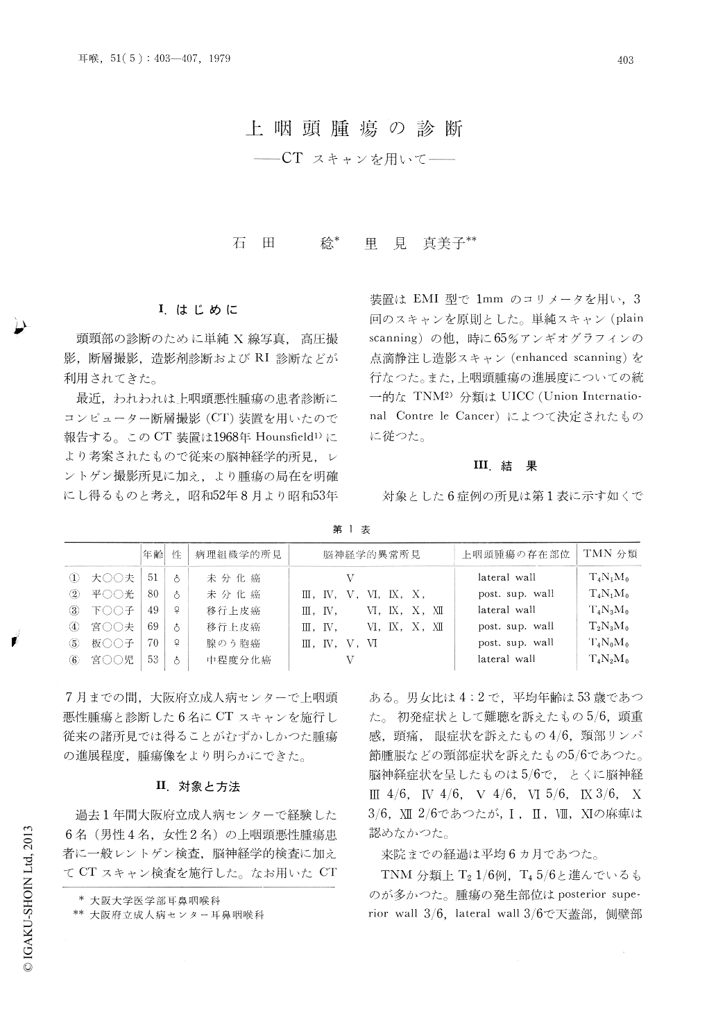
最近,われわれは上咽頭悪性腫瘍の患者診断にコンピューター断層撮影(CT)装置を用いたので報告する。このCT装置は1968年Hounsfield1)により考案されたもので従来の脳神経学的所見,レントゲン撮影所見に加え,より腫瘍の局在を明確にし得るものと考え,昭和52年8月より昭和53年7月までの間,大阪府立成人病センターで上咽頭悪性腫瘍と診断した6名にCTスキャンを施行し従来の諸所見では得ることがむずかしかつた腫瘍の進展程度,腫瘍像をより明らかにできた。
Computed tomography (CT) scanning has greatly improved the conventional diagnostic measures of nasopharyngeal cancer including X-ray and neurological examinations. The subjects studied in this report were six patients, four men and two women, averaged age of 53 years, with nasopharyngeal cancer and the patients complained initially of hearing loss, headache, and swelling of the cervical lymph nodes.
CT scanning permitted the detailed investigation of the extent of bone destruction and the direction of invasion by malignant nasopharyngeal cancer masses.
Neurological findings in four representative cases demonstrated the usefulness of the CT scanning procedure in the diagnosis and treatment of nasopharyngeal cancer.
Copyright © 1979, Igaku-Shoin Ltd. All rights reserved.