Japanese
English
- 有料閲覧
- Abstract 文献概要
- 1ページ目 Look Inside
I.はじめに
中枢神経系の海綿状血管腫は,MRIの普及とともにその報告も増え,従来考えられていたほど稀な病変ではないと思われている.しかし,脊髄髄内海綿状血管腫は極めて稀とされ1,6,10,11,14,17),脊髄髄内と脳内に多発した例はさらに稀でWoodら16)により初めて報告されて以来現在まで自験例を含めて5例の記載1,5,6,11)が見られるに過ぎない.
今回われわれは,時期を異にして発症した本邦初と思われる脊髄髄内と脳内の多発例を経験したので,若干の文献的考察を加えて報告する.
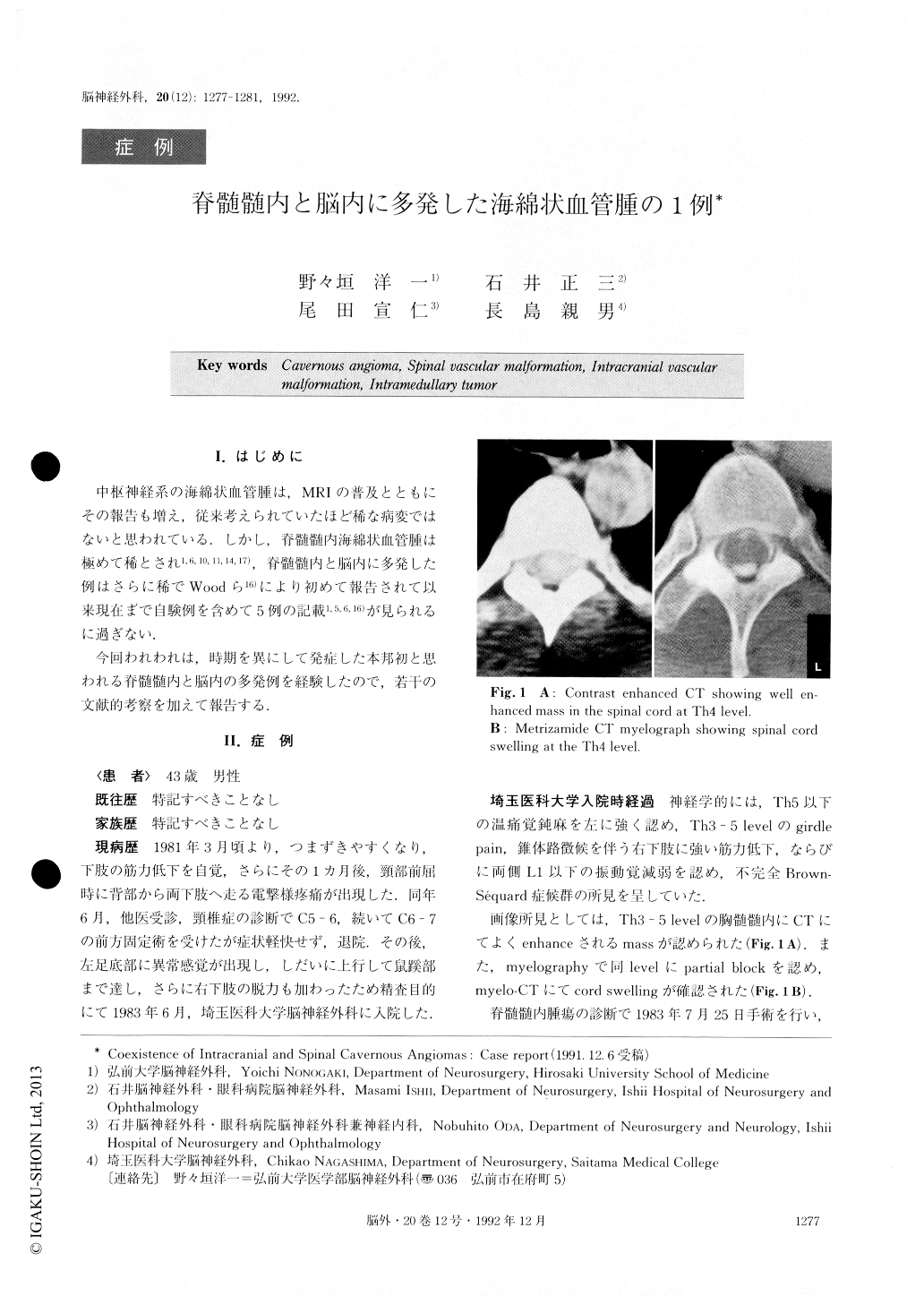
A case of a 43-year-old man with coexistence of in-tracranial and spinal cavernous angiomas is presented. The patient had a 2-year history of severe back pain in-curred by neck flexion, and he became aware of weak-ness of the right lower extremity and paresthesia of the left lower extremity. Neurological examinations at the time of the first admission demonstrated incomplete Brown-Sequard syndrome. Myelograph, myelo-CT and contrast enhanced CT showed an intramedullary mass at the Th3-Th5 level. The patient received laminectomy with total removal of the lesion. Pathological diagnosis was cavernous angioma.
Six years later, the patient complained of subacute weakness and numbness of the left upper extremity. Head CT demonstrated a high density lesion of about 2cm in diameter in the right frontal lobe. MR I showed a mixed signal intensity lesion with a marked low-intensity rim in the same area. Total extirpation of the lesion was performed. Pathological diagnosis of the intracerebral le-sion was also cavernous angioma. Intramedullary cavernous angioma is very rare. Furth-ermore, bifocal cavernous angiomas involving both the spinal cord and the brain are extremely rare, and, only 5 cases have been reported in the literature. To our know-ledge, this is the first case diagnosed by surgical speci-mens of coexisting intramedullary and intracerebral lesions.

Copyright © 1992, Igaku-Shoin Ltd. All rights reserved.


