Japanese
English
- 有料閲覧
PPV購入案内
1,320円 (1,200円+税10%) こちらは、1079ページから1083ページの文献です。購入には医書.jp本会員登録が必要です。
会員登録完了後に改めて上記「購入サイトへ」を選択してください。
または「購入サイトへ」選択後に「購入ログイン」、「新規会員登録」の順に進んでください。
会員登録について詳しくはをご確認ください。
- Abstract 文献概要
- 1ページ目 Look Inside
I.はじめに
頭蓋内に発生する海綿状血管腫は比較的まれな疾患である.頭蓋内脳外としては中頭蓋窩に発生するものが知られているが,それ以外の部位のものはさらにまれである.われわれはこれまで報告のない大脳鎌に発生した海綿状血管腫を経験したので若干の文献的考察を加え報告する.
Abstract
A first case of cavernous angioma of falx cerebri is reported.
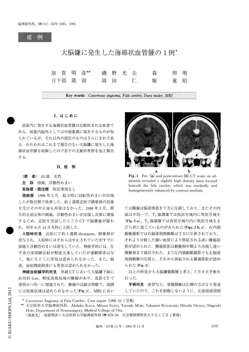
A 62-year-old woman who had a history of intermit-tent headache and dizziness was admitted to our hospit-al. On admission she had no neurological deficit, but CT scan showed a slightly high density tumor located beneath the falx cerebri. This was markedly and homogeneously enhanced by contrast medium. MRI showed a tumor with low intensity in Tl-weighted im-age and high intensity speckled with low intensity in T2-weighted image. Angiogram revealed a faint tumor-stain fed by the bilateral pericallosal arteries at middle arterial to late venous phases.
With the tumor attached to the lower edge, the falx was totally removed through a left front-parietal cra-niotomy. Histologically the tumor was diagnosed as cavernous angioma and thought to have originated from the dura mater of the falx.
A search in the literature revealed that only 7 cases of extracerebral cavernous angiomas excluding ones in the middle cranial fossa have been previously reported. Five of them were located at the tentorium cerebelli and two at the convexity. The MRI finding such as speckled mixed intensity may reflect the vascular lu-mens or their thromboses in the tumor. Angiographic finding such as faint tumor stain at middle arterial to late venous phases due to slow blood flow in the tumor are thought to be specific to intracranial cavernous angiomas. These findings are of particular importance in differentiating cavernous angiomas from menin-giomas.
Copyright © 1991, Igaku-Shoin Ltd. All rights reserved.