Japanese
English
- 有料閲覧
- Abstract 文献概要
- 1ページ目 Look Inside
I.はじめに
CT, MRIなどの画像診断の進歩により,中枢神経系の海綿状血管腫(CA)の診断は容易となった.血管性奇形の1つであり髄内に好発するCAが内耳道・小脳橋角部に発生することは稀であり,本邦における報告は見あたらない.われわれは小脳橋角部CAの1例を経験したので報告し,文献報告17例に本症例を加えた18例を対象に本症の特徴について検討する.
A rare case of cavernous angioma (CA) in the cerebel-lopontine angle (CPA) is reported.
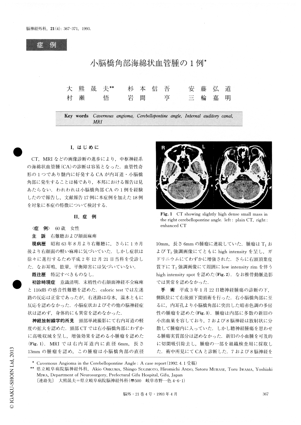
A 60-year-old female suffered from a right progressive sensorineural hearing loss and a successive right facial paresis over 2 years. A small mass was detected in her right CPA on CT scans. Both T1- and T2-weighted MR images demonstrated an intracanalicular lesion protrud-ing into the CPA as being hyperintense. A small red col-ored lobulated tumor involving the 7th and 8th cranial nerves was found in the CPA through the suboccipital approach. The tumor contained multiple small hemato-mas in various stages. Biopsy with evacuation of these hematomas was selected to avoid damaging the cranial nerves. Histological examination of the specimen dis-closed it as CA. Postoperatively her facial paresis im-proved slightly, but her hearing loss remained un-changed.
Discussions were carried out concerning clinical andneuroradiological features of CA in the internal auditory canal and CPA. The present case and a previously re-ported 17 cases were the subjects under discussion.

Copyright © 1993, Igaku-Shoin Ltd. All rights reserved.


