Japanese
English
- 有料閲覧
- Abstract 文献概要
- 1ページ目 Look Inside
I.はじめに
脳幹部の血管腫に対する直達手術の報告は稀である1-6,8-10,14,19).特に中脳においてはこれまでに数例を数えるのみである1,2,4,6,10).しかし,MRIなどの画像診断および手術手技の進歩により12),これまで直達手術が不可能と考えられていた脳幹部病変に対しても手術適応となるものが今後増加してくるものと考えられる.今回,われわれは出血発作を繰り返し,次第に神経症状の増悪を認めた中脳の大きな海綿状血管腫に対して,3回の手術により全摘出を完遂し得た症例を経験したので,若干の文献的考察を加え報告する.
The authors presented a case of microsurgically re-moved large cavernous angioma in the midbrain.
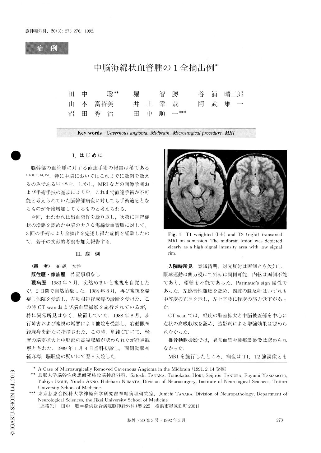
A 46-year old woman was admitted to our service with bilateral nuclear oculomotor pareses and mild tet-raparesis. MRI revealed a large round well-circumscribed high signal-intensity lesion on both Ti and T2 weighted image with low signal rim in the mid-brain. This lesion was diagnosed preoperatively as a cavernous angioma.
The operation was performed in three steps with in-termittent hemorrhages by interhemispheric transcallo-sal-hippocampal commissure - velum interpositum-third ventricular approach and subsequent two infratentorial supracerebellar approaches and finally complete remov-al was performed. Histological examination of the sur-gical specimen revealed as a cavernous angioma having abnormal vessels with honeycomb appearance. The pa-tient survived, although remains moderately disabled 6 months after the last Operation.
Copyright © 1992, Igaku-Shoin Ltd. All rights reserved.