Japanese
English
- 有料閲覧
PPV購入案内
1,320円 (1,200円+税10%) こちらは、979ページから983ページの文献です。購入には医書.jp本会員登録が必要です。
会員登録完了後に改めて上記「購入サイトへ」を選択してください。
または「購入サイトへ」選択後に「購入ログイン」、「新規会員登録」の順に進んでください。
会員登録について詳しくはをご確認ください。
- Abstract 文献概要
- 1ページ目 Look Inside
I.はじめに
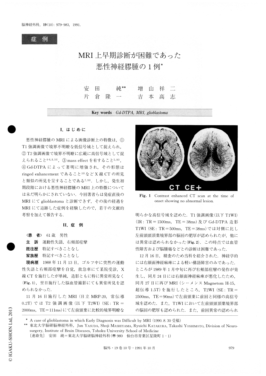
悪性神経膠腫のMRIによる画像診断上の特徴は,①T1強調画像で境界不明瞭な低信号域として捉えられ,②T2強調画像で境界不明瞭に広範に高信号域として捉えられること4-6,8,10),③mass effectを有すること5,10),④Gd-DTPAによって著明に増強され,その形態はringed enhancementであること10)などX線CTの所見と類似の所見を呈することである7,10).しかし,発生初期段階における悪性神経膠腫のMRI上の特徴については未だ明らかにされていない.今回著者らは発症直後のMRIにてglioblastomaと診断できず,その後の経過をMRIにて追跡した症例を経験したので,若干の文献的考察を加えて報告する.
Abstract
The authors report a case of glioblastoma in which MR images with Gd-DTPA enhancement changed rapidly during the early stage.
A 61 year-old male presented with sudden right fa-cial spasm and dysarthria. However, both a plain and an enhanced CT failed to demonstrate any abnormal le-sions. On the other hand, T2 weighted MR image re-vealed a well circumscribed high intensity lesion in the left frontal lobe without mass effect. This lesion couldnot be differentiated from cerebral infarction, since no contrast enhanced lesion was able to be observed in T1 weighted MR image with Gd-DTPA. His symptoms gradually became aggravated and at 3 months from the onset, MR image with Gd-DTPA disclosed a small en-hanced lesion in the left frontal lobe neat the cortical surface. After 6 months from the onset, he suffered from right hemiparesis and motor aphasia. The MR im-age with Gd-DTPA at this time showed a large en-hanced lesion in the left frontal lobe with mass effect. He was admitted to our hospital, and subtotal removal of the tumor and intraoperative radiation was carried out. The patient did well postoperatiyely without addi-tional neurological deficit, and then he received addi-tional radiation therapy.
It should be noted that Gd-DTPA enhanced MR im-age might fail to reveal the lesion of glioblastoma in its early stage, while Ti weighted image discloses only the gyral swelling. This lesion couldnot be differentiated from cerebral infarction, since no contrast enhanced lesion was able to be observed in T1 weighted MR image with Gd-DTPA. His symptoms gradually became aggravated and at 3 months from the onset, MR image with Gd-DTPA disclosed a small en-hanced lesion in the left frontal lobe neat the cortical surface. After 6 months from the onset, he suffered from right hemiparesis and motor aphasia. The MR im-age with Gd-DTPA at this time showed a large en-hanced lesion in the left frontal lobe with mass effect. He was admitted to our hospital, and subtotal removal of the tumor and intraoperative radiation was carried out. The patient did well postoperatiyely without addi-tional neurological deficit, and then he received addi-tional radiation therapy.
It should be noted that Gd-DTPA enhanced MR im-age might fail to reveal the lesion of glioblastoma in its early stage, while Ti weighted image discloses only the gyral swelling.
Copyright © 1991, Igaku-Shoin Ltd. All rights reserved.