Japanese
English
- 有料閲覧
PPV購入案内
1,320円 (1,200円+税10%) こちらは、449ページから455ページの文献です。購入には医書.jp本会員登録が必要です。
会員登録完了後に改めて上記「購入サイトへ」を選択してください。
または「購入サイトへ」選択後に「購入ログイン」、「新規会員登録」の順に進んでください。
会員登録について詳しくはをご確認ください。
- Abstract 文献概要
- 1ページ目 Look Inside
I.はじめに
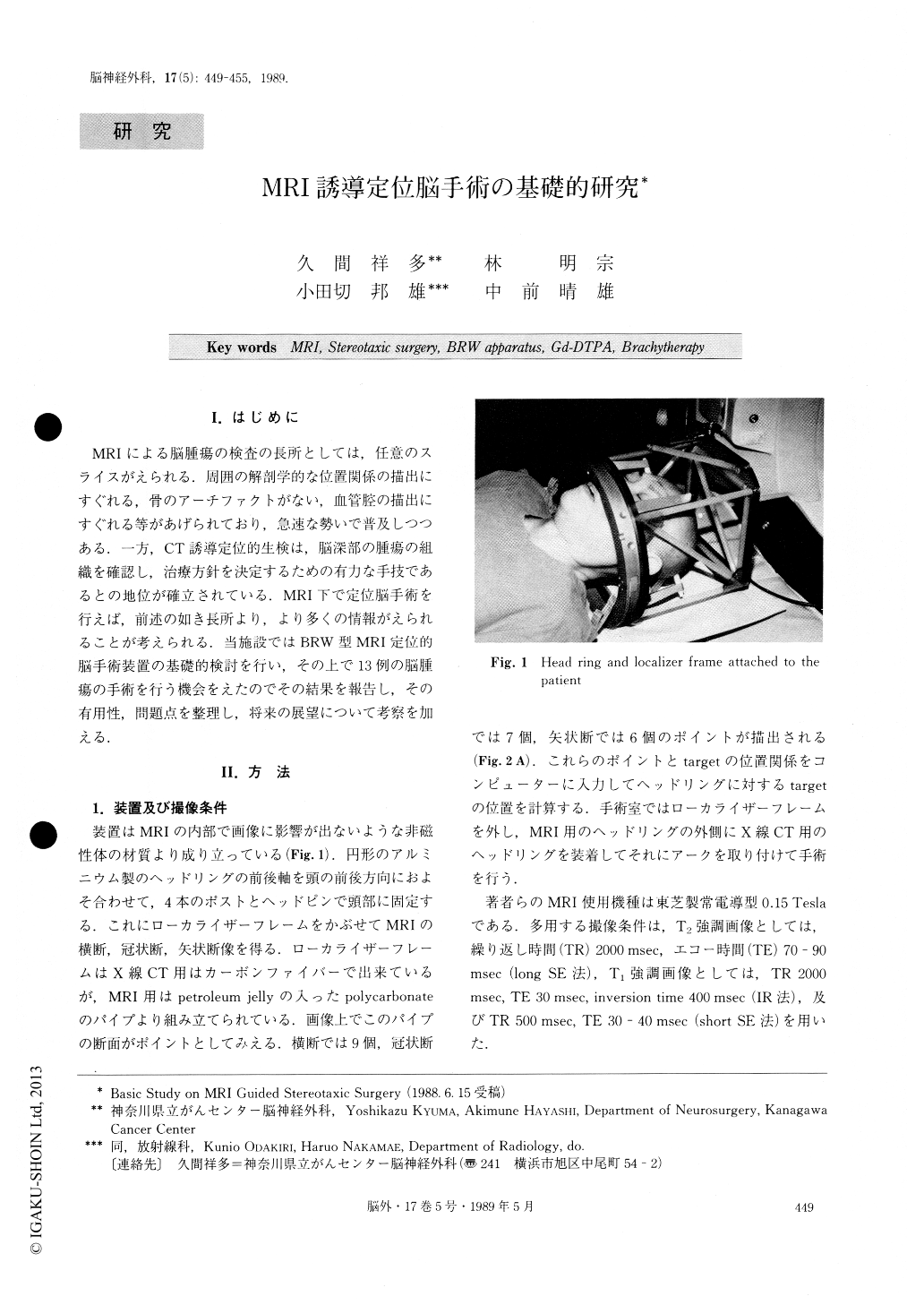
MRIによる脳腫瘍の検査の長所としては,任意のスライスがえられる.周囲の解剖学的な位置関係の描出にすぐれる,骨のアーチファクトがない,血管腔の描出にすぐれる等があげられており,急速な勢いで普及しつつある.一方,CT誘導定位的生検は,脳深部の腫瘍の組織を確認し,治療方針を決定するための有力な手技であるとの地位が確立されている.MRI下で定位脳手術を行えば,前述の如き長所より,より多くの情報がえられることが考えられる.当施設ではBRW型MRI定位的脳手術装置の基礎的検討を行い,その上で13例の脳腫瘍の手術を行う機会をえたのでその結果を報告し,その有用性,問題点を整理し,将来の展望について考察を加える.
In 1987, we started MRI guided stereotaxic surgery using BRW-MRI apparatus. As distortion of image had been noted around the periphery of MRI due to the characteristic static magnetic field, we examined accuracy of data before clinical application. The MRI machine was a MRT-15A, 0.15 Tesla resistive type made by Toshiba. The stereotaxic apparatus consisted of a head ring, which was fixed to the patient's head, a cubical localizer frame which was attached to the head ring for the scanning phase, and a programmed compu-ter.
Copyright © 1989, Igaku-Shoin Ltd. All rights reserved.