Japanese
English
- 有料閲覧
PPV購入案内
1,320円 (1,200円+税10%) こちらは、415ページから424ページの文献です。購入には医書.jp本会員登録が必要です。
会員登録完了後に改めて上記「購入サイトへ」を選択してください。
または「購入サイトへ」選択後に「購入ログイン」、「新規会員登録」の順に進んでください。
会員登録について詳しくはをご確認ください。
- Abstract 文献概要
- 1ページ目 Look Inside
- 参考文献 Reference
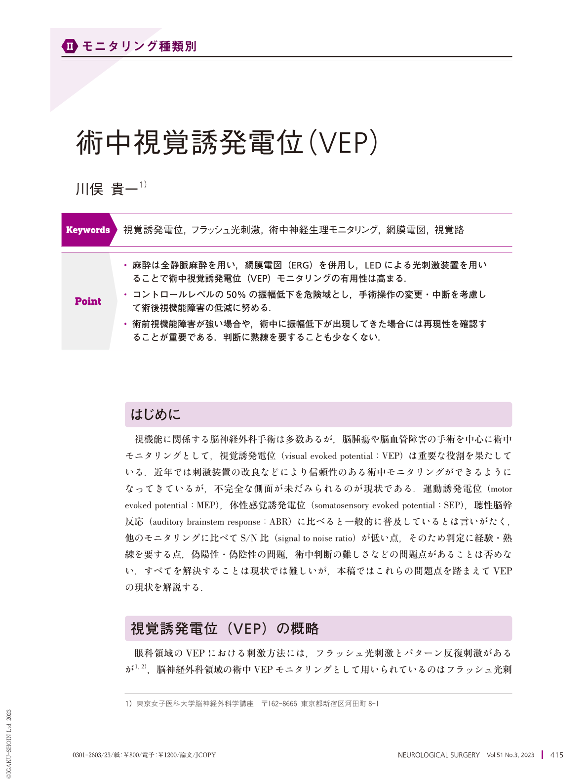
Point
・麻酔は全静脈麻酔を用い,網膜電図(ERG)を併用し,LEDによる光刺激装置を用いることで術中視覚誘発電位(VEP)モニタリングの有用性は高まる.
・コントロールレベルの50%の振幅低下を危険域とし,手術操作の変更・中断を考慮して術後視機能障害の低減に努める.
・術前視機能障害が強い場合や,術中に振幅低下が出現してきた場合には再現性を確認することが重要である.判断に熟練を要することも少なくない.
In neurosurgery, the intraoperative visual evoked potential(VEP)has recently been used for the management of anterior skull base and parasellar tumors related to the optic pathways to prevent postoperative visual complications. We used light emitting diode photo-stimulation thin pad and stimulator(Unique Medical, Japan). We also recorded the electroretinogram(ERG)simultaneously to exclude technical errors. VEP is defined as an amplitude between the maximum positive wave at 100 ms(P100)and the prior negative wave(N75).
In intraoperative VEP monitoring, reproducibility of VEP should be ascertained, particularly in patients with preoperative advanced visual impairment and an intraoperative diminished amplitude. Furthermore, a 50% reduction in the amplitude is critical. In such cases, we should consider suspending or changing surgical manipulation.
We have not clearly verified the relationship between the absolute intraoperative VEP value and postoperative visual function. Mild peripheral visual field defects cannot be detected in the present intraoperative VEP system. However, intraoperative VEP with ERG monitoring can serve as a real-time warning to guide surgeons to avoid postoperative visual impairment. We should comprehend the principles, characteristics, disadvantages, and limitations of intraoperative VEP monitoring for reliable and effective utilization.
Copyright © 2023, Igaku-Shoin Ltd. All rights reserved.