Japanese
English
- 有料閲覧
PPV購入案内
1,320円 (1,200円+税10%) こちらは、611ページから617ページの文献です。購入には医書.jp本会員登録が必要です。
会員登録完了後に改めて上記「購入サイトへ」を選択してください。
または「購入サイトへ」選択後に「購入ログイン」、「新規会員登録」の順に進んでください。
会員登録について詳しくはをご確認ください。
- Abstract 文献概要
- 1ページ目 Look Inside
- 参考文献 Reference
Ⅰ.はじめに
現代の脳神経外科手術において,手術用顕微鏡や神経内視鏡は必要不可欠な光学機器である5).近年,明るく高倍率で高精細な画像が得られ手術記録も可能な体外視鏡(exoscope)が耳鼻咽喉科や婦人科領域での手術・手技で使用され13,14),脳神経外科領域でも使用されはじめている1,2,5,6,12).本稿ではexoscope systemの概要と脳神経外科手術における使用報告例をreviewした.さらにexoscopeとtubular retractor,diffusion tensor imaging tractography,脳神経外科手術用ナビゲーションシステムを併用した脳深部病変へのアプローチについても紹介する.
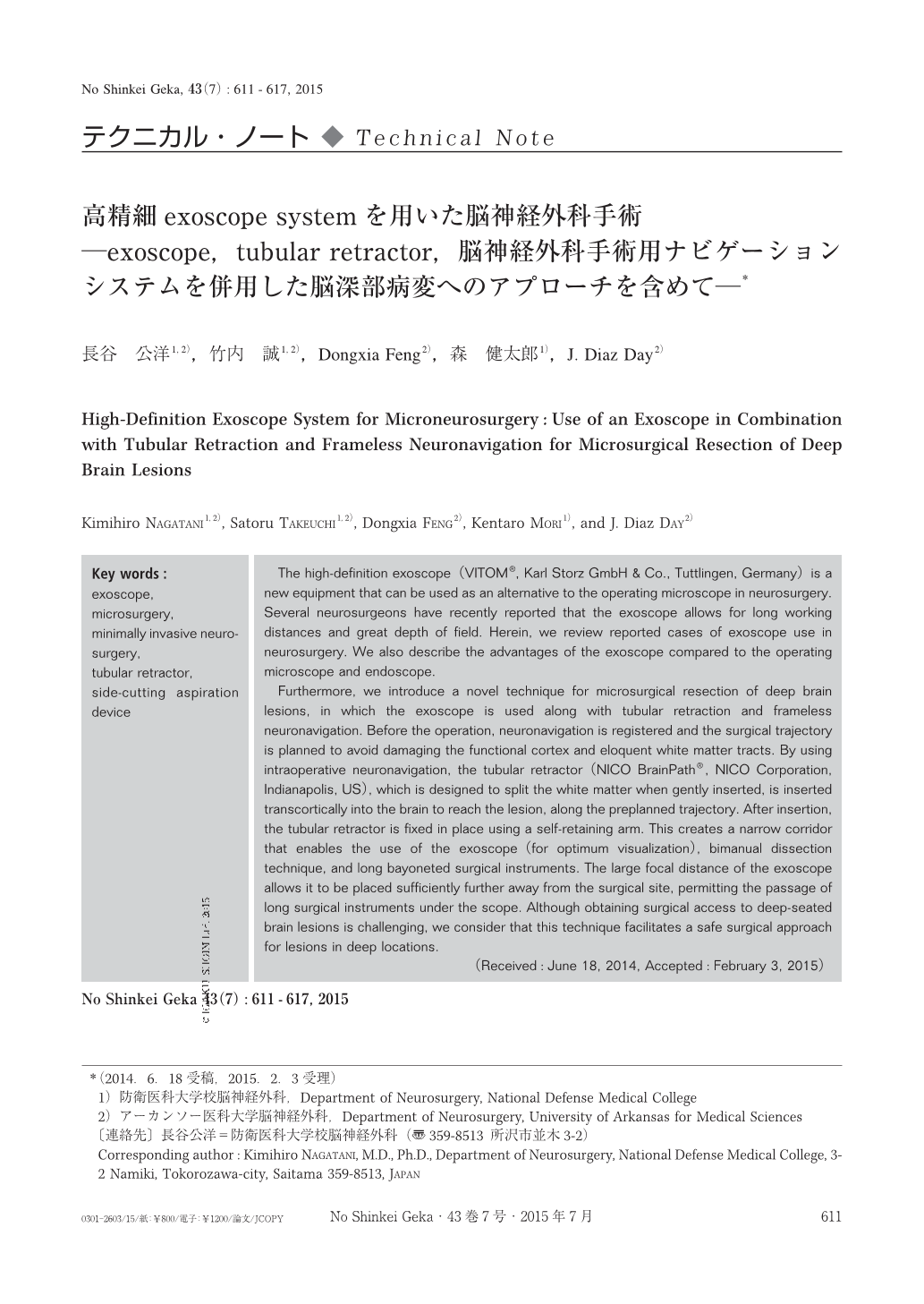
The high-definition exoscope(VITOM®, Karl Storz GmbH & Co., Tuttlingen, Germany)is a new equipment that can be used as an alternative to the operating microscope in neurosurgery. Several neurosurgeons have recently reported that the exoscope allows for long working distances and great depth of field. Herein, we review reported cases of exoscope use in neurosurgery. We also describe the advantages of the exoscope compared to the operating microscope and endoscope.
Furthermore, we introduce a novel technique for microsurgical resection of deep brain lesions, in which the exoscope is used along with tubular retraction and frameless neuronavigation. Before the operation, neuronavigation is registered and the surgical trajectory is planned to avoid damaging the functional cortex and eloquent white matter tracts. By using intraoperative neuronavigation, the tubular retractor(NICO BrainPath®, NICO Corporation, Indianapolis, US), which is designed to split the white matter when gently inserted, is inserted transcortically into the brain to reach the lesion, along the preplanned trajectory. After insertion, the tubular retractor is fixed in place using a self-retaining arm. This creates a narrow corridor that enables the use of the exoscope(for optimum visualization), bimanual dissection technique, and long bayoneted surgical instruments. The large focal distance of the exoscope allows it to be placed sufficiently further away from the surgical site, permitting the passage of long surgical instruments under the scope. Although obtaining surgical access to deep-seated brain lesions is challenging, we consider that this technique facilitates a safe surgical approach for lesions in deep locations.
Copyright © 2015, Igaku-Shoin Ltd. All rights reserved.