Japanese
English
症例
頭蓋内骨軟骨腫の1例—特にそのMRI像について
A Case of Intracranial Osteochondroma: Its MR images
畑山 徹
1
,
関谷 徹治
1
,
鈴木 重晴
1
,
岩淵 隆
1
Toru HATAYAMA
1
,
Tetsuji SEKIYA
1
,
Shigeharu SUZUKI
1
,
Takashi IWABUCHI
1
1弘前大学脳神経外科
1Department of Neurosurgery, Hirosaki University School of Medicine
キーワード:
Osteochondroma
,
CT
,
MRI
Keyword:
Osteochondroma
,
CT
,
MRI
pp.1063-1066
発行日 1989年11月10日
Published Date 1989/11/10
DOI https://doi.org/10.11477/mf.1436202917
- 有料閲覧
PPV購入案内
1,320円 (1,200円+税10%) こちらは、1063ページから1066ページの文献です。
PPV(ペイ・パー・ビュー)とは、論文(記事)単位で購入・閲覧ができるサービスです。
購入には医書.jp本会員登録が必要です。
会員登録完了後に改めて上記「購入サイトへ」を選択してください。
または「購入サイトへ」選択後に「購入ログイン」、「新規会員登録」の順に進んでください。
会員登録について詳しくはをご確認ください。
購入には医書.jp本会員登録が必要です。
会員登録完了後に改めて上記「購入サイトへ」を選択してください。
または「購入サイトへ」選択後に「購入ログイン」、「新規会員登録」の順に進んでください。
会員登録について詳しくはをご確認ください。
- Abstract 文献概要
- 1ページ目 Look Inside
I.はじめに
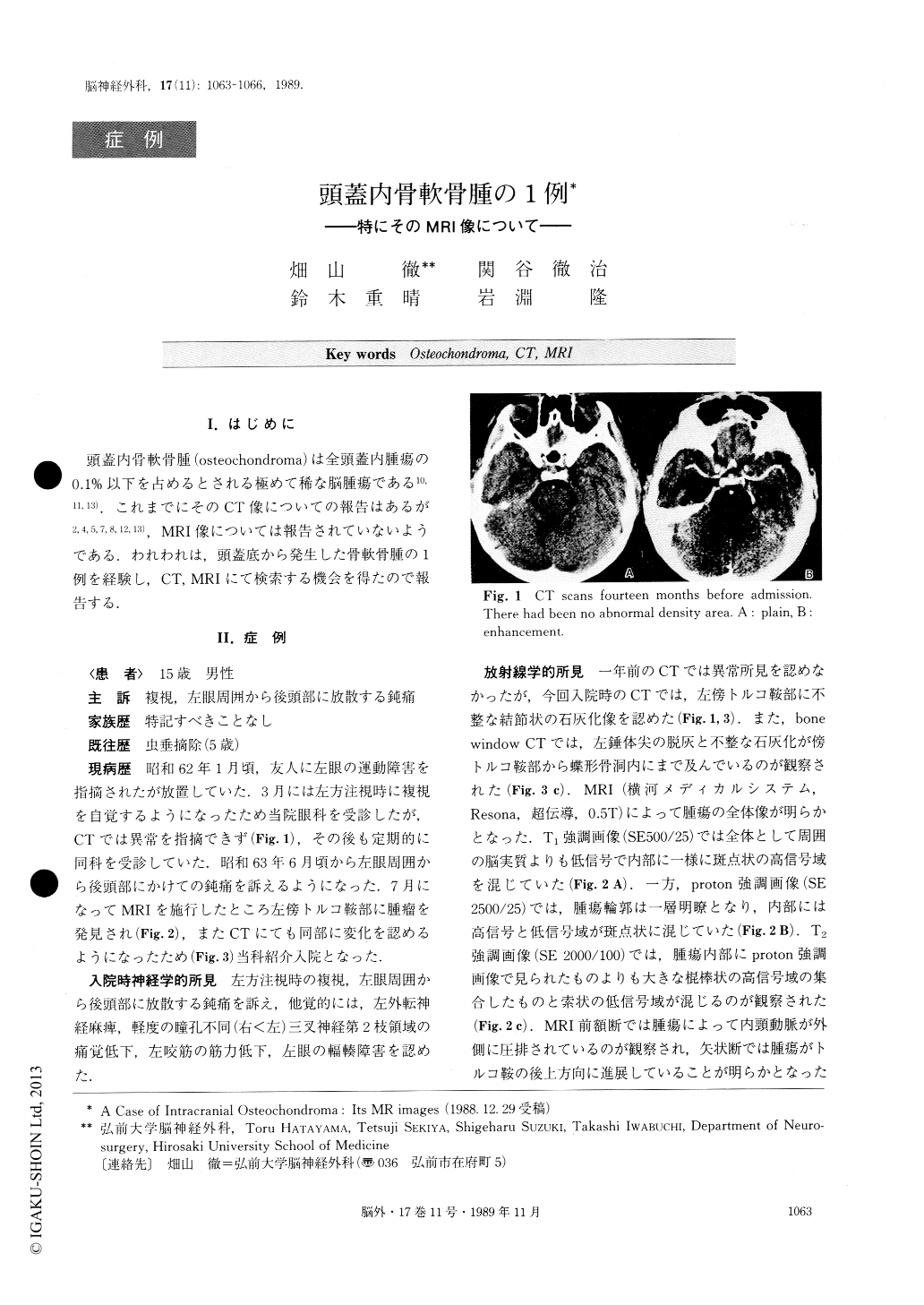
頭蓋内骨軟骨腫(osteochondroma)は全頭蓋内腫瘍の0.1%以下を占めるとされる極めて稀な脳腫瘍である10,11,13).これまでにそのCT像についての報告はあるが2,4,5,7,8,12,13),MRI像については報告されていないようである.われわれは,頭蓋底から発生した骨軟骨腫の1例を経験し,CT,MRIにて検索する機会を得たので報告する.
A 15-year-old boy was referred to our department due to left abducens palsy and trigeminal neuralgia. CT scans that had been taken fourteen months before this referral failed to disclose any abnormality. CT scans taken on admission, however, demonstrated an irregu-lar calcified lesion in the left parasellar region. This im-plied that some ossifying process had been progressing during these fourteen months.
MR images clearly demonstrated tumor contour and it was shown that high and low intensity spots were evenly scattered within the tumor.
Copyright © 1989, Igaku-Shoin Ltd. All rights reserved.