Japanese
English
- 有料閲覧
- Abstract 文献概要
- 1ページ目 Look Inside
I.はじめに
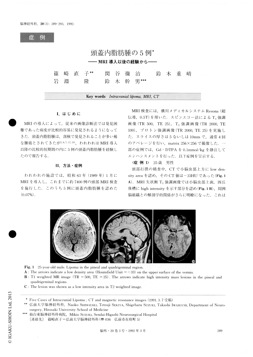
MRIの導人によって,従来の画像診断法では発見困難であった病変が比較的容易に発見されるようになってきた.頭蓋内脂肪腫は,剖検で発見されることが多い稀な腫瘍とされてきたが2,6,7,12,15),われわれはMRI導人以降の比較的知期間の内に5例の頭蓋内脂肪腫を経験したので報告する.
We encountered five cases of intracranial lipoma af-ter introduction of MRI. They were located in the quadrigeminal plate, interpeduncular fossa, pineal re-gion and two of them were found in the cerehellopon-tine angle, (although intracranial lipoma in this location has been reported to be extremely rare). MRI can pre-cisely locate a small lesion that would be overlooked by CT scans. Operative treatment was performed in two symptomatic cases (CP angle and pineal lesions) and the tumors were subtotally resected. The symptoms of the patients disappeared postoperatively. This indicated that even subtotal removal can alleviate the symptoms of intracranial lipomas and that favorable results can be obtained.

Copyright © 1992, Igaku-Shoin Ltd. All rights reserved.


