Japanese
English
- 有料閲覧
PPV購入案内
1,320円 (1,200円+税10%) こちらは、987ページから992ページの文献です。購入には医書.jp本会員登録が必要です。
会員登録完了後に改めて上記「購入サイトへ」を選択してください。
または「購入サイトへ」選択後に「購入ログイン」、「新規会員登録」の順に進んでください。
会員登録について詳しくはをご確認ください。
- Abstract 文献概要
- 1ページ目 Look Inside
I.はじめに
脳幹内で電気刺激を行い頭蓋外の各支配筋からcom—pound muscle action potentials(以下,CMAP)を記録することによって,脳神経核のmappingを行うことができる2,5,9-13).しかし,これまでの報告では,顔面神経核と下位脳神経核のmappingの報告は多いが,眼球運動神経核の同定の報告は極めて乏しい5,9,10,13).中脳手術において,眼球運動神経核やその髄内路をmappingすることの意義は大きいと考えられるにも関わらずmap—pingの報告が乏しい原因の一つは,外眼筋に直接電極を設置することが困難なことにあると考えられる.
われわれは,眼球運動神経系の術中モニタリングをルーチン・ベースで行ってきているが11,12),このモニタリング法のより簡便な実施方法,すなわち,市販の電極を用いて眼科医がいない状況下でも実施できるような方法も同時に模索してきた.その結果,より簡便な術中モニタリング法を開発することができたので報告する.
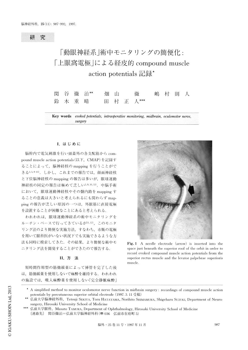
We report a simple and easily-applicable method to intraoperatively monitor the oculomotor nerve and ocu-lomotor nucleus in the brainstem. A needle electrode is inserted into the space just beneath the superior roof of the orbit. With this superior orbital electrode, evoked compound muscle action potentials from the superior rectus and the levator palpebrae superioris muscles were recorded. The nuclei for these two muscles are lo-cated caudally in the midline of the midbrain. The re-sponses from the superior orbital electrode, therefore, give the surgeons useful intraoperative guidance during surgery for pineal region tumors and midbrain gliomas.
Copyright © 1997, Igaku-Shoin Ltd. All rights reserved.