Japanese
English
- 有料閲覧
PPV購入案内
1,320円 (1,200円+税10%) こちらは、947ページから951ページの文献です。購入には医書.jp本会員登録が必要です。
会員登録完了後に改めて上記「購入サイトへ」を選択してください。
または「購入サイトへ」選択後に「購入ログイン」、「新規会員登録」の順に進んでください。
会員登録について詳しくはをご確認ください。
- Abstract 文献概要
- 1ページ目 Look Inside
I.はじめに
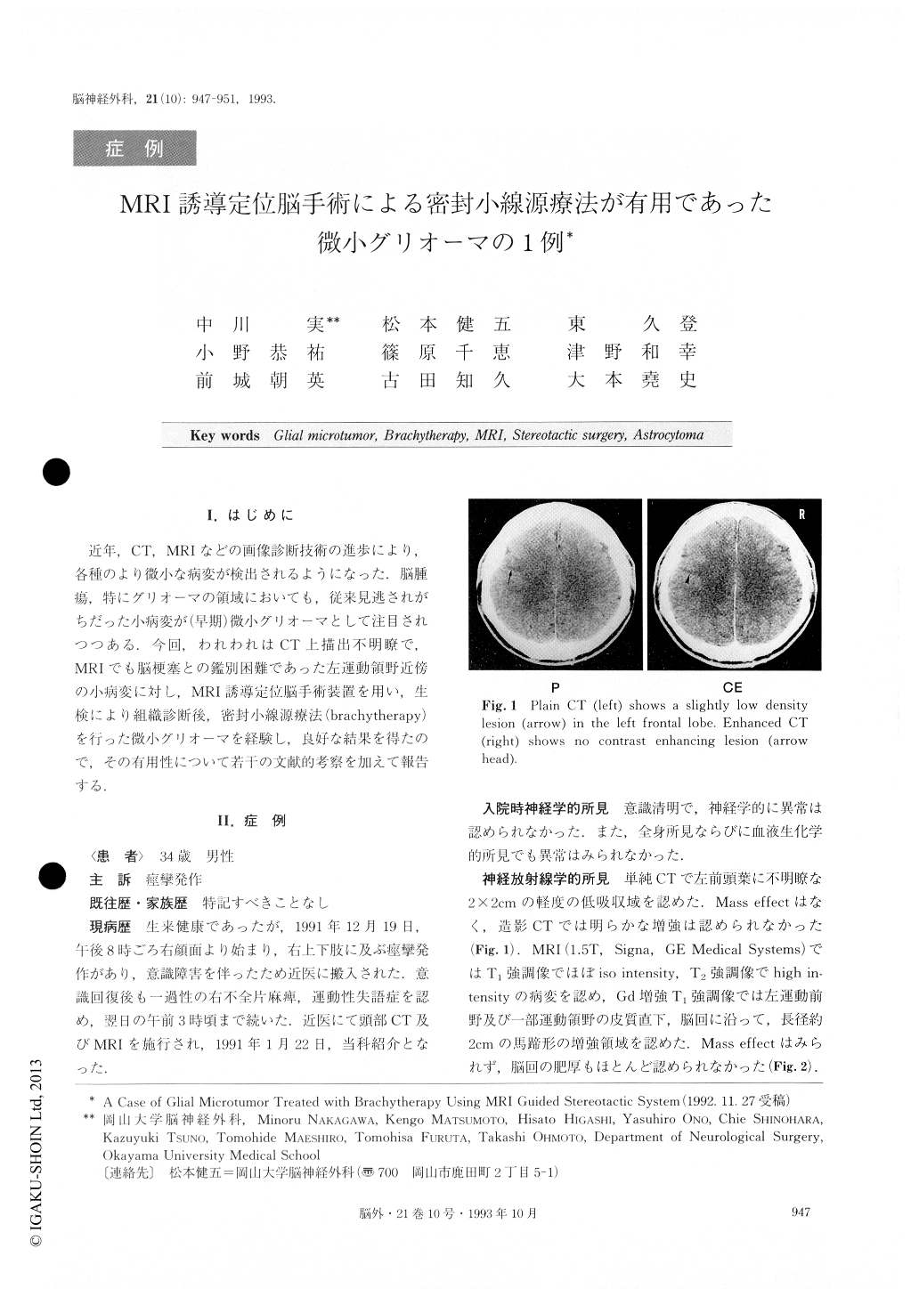
近年,CT,MRIなどの画像診断技術の進歩により,各種のより微小な病変が検出されるようになった.脳腫瘍,特にグリオーマの領域においても,従来見逃されがちだった小病変が(早期)微小グリオーマとして注目されつつある.今回,われわれはCTL描出不明瞭で,MRIでも脳梗塞との鑑別困難であった左運動領野近傍の小病変に対し,MRI誘導定位脳手術装概を用い,生検により組織診断後,密封小線源療法(brachytherapy)を行った微小グリオーマを経験し,良好な結果を得たので,その有用性について若トの文献的考察を加えて報告する.
A 34 year old male suffered from convulsion on his right side with loss of consciousness. Neither plain nor enhanced CT could clearly demonstrate a lesion. Howev-er, a Gd-DTPA enhanced Ti weighted image (T1WI) de-monstrated an enhancing lesion (2cm in diameter) in the left frontal lobe including the motor cortex. This lesion was not identified on plain T1WI. A T2, weighted image (T2WI) showed a high intensity lesion without mass effect slightly larger than the enhanced area on "HIW.This lesion was suspected to be a glial microtumor, but, because it was difficult to differentiate it from a cerebral infarction, it was confirmed to be a neoplasm by means of an MRI guided stereotactic biopsy. The histological dia-gnosis of the frozen section was a grade 2 astrocytoma.
Subsequently brachytherapy with seeds was per-formed, and about 80Gy of interstitial irradiation was given to the edge of the enhanced area on MRI. The minimum dose in the high intensity area on T2WI was about 50Gy. The patient was discharged with no neuro-logical deficits. The Gd-DTPA enhanced lesion decreased in size six months after the treatment.
MRI guided stereotactic system makes it possible to target a high intensity lesion on T,WI in brachytherapy. Brachytherapy using this system is considered to be use-ful in the treatment of glial microtumor in an area in which it is difficult to identify it on CT.
Copyright © 1993, Igaku-Shoin Ltd. All rights reserved.