Japanese
English
- 有料閲覧
- Abstract 文献概要
- 1ページ目 Look Inside
I.はじめに
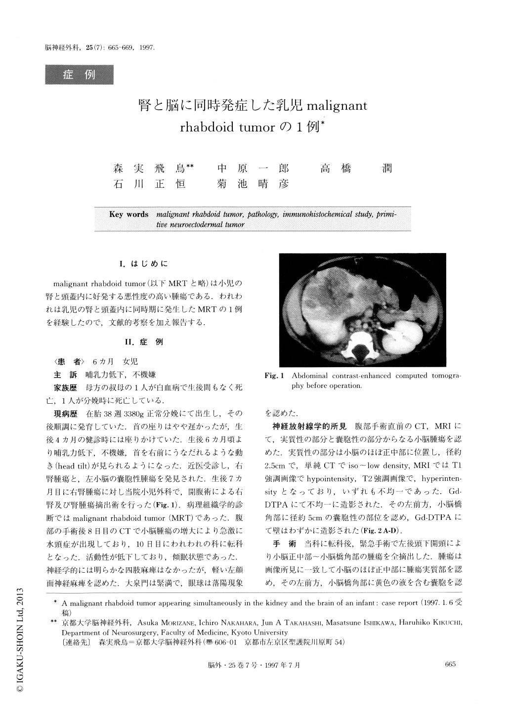
malignant rhabdoid tumor(以下MRTと略)は小児の腎と頭蓋内に好発する悪性度の高い腫瘍である.われわれは乳児の腎と頭蓋内に同時期に発生したMRTの1例を経験したので,文献的考察を加え報告する.
A 6-month-old female was admitted to the hospital with bad temper and decreased sucking power. CT scans revealed tumors in her right kidney and left cere-bellum. The patient underwent right radical nephrec-tomy to excise the kidney tumor. The pathological di-agnosis was malignant rhabdoid tumor (MRT). Seven days later, the patient underwent left suboccipital cra-niectomy for total excision of the cerebellar tumor. The cerebellar tumor existed extraaxially, and consisted of a solid mass lesion and a cystic lesion. Histological ex-amination revealed that it was also a malignant rhab-doid tumor. A follow-up CT, 1.5 months after surgery, revealed a recurrence of the kidney tumor and metasta-sis to the chest wall and lung. The patient received 16.9Gy radiotherapy to the abdominal tumor and che-motherapy with etoposide, carboplatin, and ifosfamide. However, she died of respiratory insufficiency 4 months after surgery, though neither recurrence nor metastasis was found in the brain. Nor was there evi-dence of leptomeningeal dissemination.
MRT is a highly malignant tumor that occurs most frequently in the kidney. However, it can also occur in other tissues, including the brain. This tumor occurs most commonly in children under 2 years of age. There is a 3: 2 male predominance. The median length of overall survival of MRT in the brain is 6 months. MRT contains nests or sheets of rhabdoid cells. A typical rhabdoid cell has an eccentric round nucleus with a prominent nucleolus and a plump cell body. MRT is composed entirely or partly of rhabdoid cells. Many MRTs have other components, such as PNET areas, mesenchymal area, and epithelial areas. For this reason, they are sometimes called atypical teratoid/rhab-doid tumors. MRTs in the brain contain fewer rhab-doid cell areas than MRT in the kidney. This makes di-agnosing MRT in the brain more difficult. A careful search of the entire specimen for variations in pattern and cell type, along with application of immunohis-tochemical methods is the most useful method of obtaining a diagnosis.
In our case, the cerebellar tumor consisted of rhab-doid cell areas, mesenchymal areas, and PNET areas. The cerebellar tumor contained fewer rhabdoid cell areas than the kidney tumor. However, the rhabdoid cell areas in the cerebellar tumor were almost the same as those in the kidney tumor. Furthermore, immunohis-tochemical staining was positive for vimentin and kera-tin in the rhabdoid cell areas. Therefore, we were able to make a diagnosis of MRT.
It is possible that some of the previously reported cases diagnosed as CNS PNET were actually MRT in the brain, especially if the cases were associated with MRT in the kidney.
Copyright © 1997, Igaku-Shoin Ltd. All rights reserved.