Japanese
English
- 有料閲覧
PPV購入案内
1,320円 (1,200円+税10%) こちらは、1487ページから1491ページの文献です。購入には医書.jp本会員登録が必要です。
会員登録完了後に改めて上記「購入サイトへ」を選択してください。
または「購入サイトへ」選択後に「購入ログイン」、「新規会員登録」の順に進んでください。
会員登録について詳しくはをご確認ください。
- Abstract 文献概要
- 1ページ目 Look Inside
I.はじめに
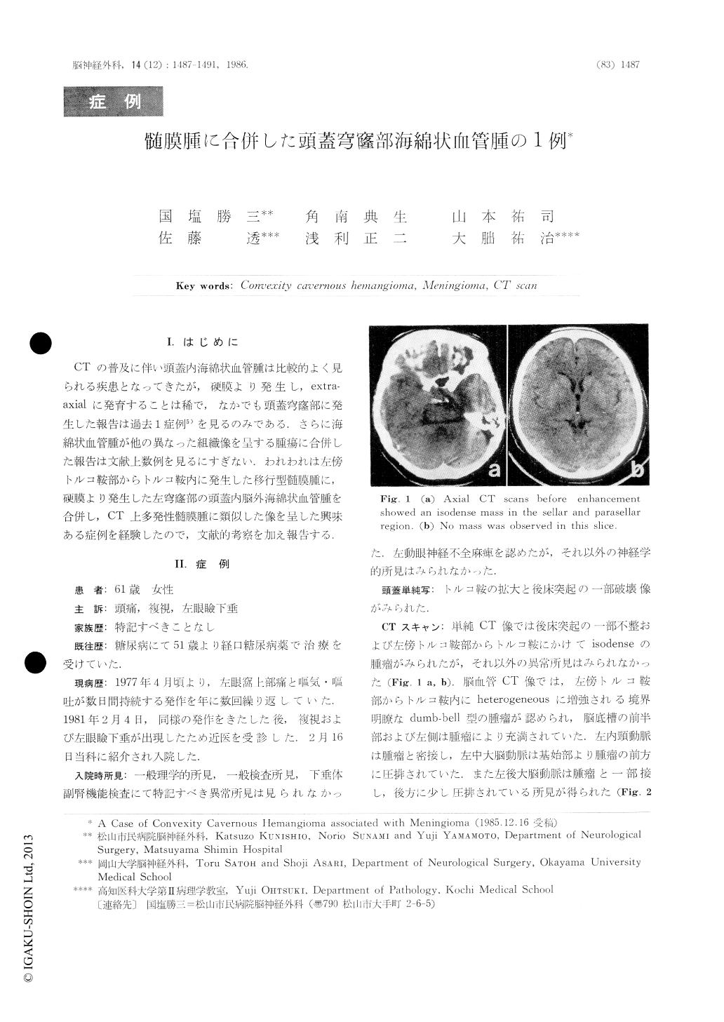
CTの普及に伴い頭蓋内海綿状血管腫は比較的よく見られる疾患となってきたが,硬膜より発生し,extra—axialに発育することは稀で,なかでも頭蓋穹窿部に発生した報告は過去1症例5)を見るのみである.さらに海綿状血管腫が他の異なった組織像を呈する腫瘍に合併した報告は文献上数例を見るにすぎない.われわれは左傍トルコ鞍部からトルコ鞍内に発生した移行型髄膜腫に,硬膜より発生した左穹窿部の頭蓋内脳外海綿状血管腫を合併し,CT上多発性髄膜腫に類似した像を呈した興味ある症例を経験したので,文献的考察を加え報告する.
A case of convexity cavernous hemangioma as-sociated with seller meningioma with parasellar ex-tension is presented.
A 61-year-old female who had complained of left blepharoptosis and diplopia was admitted to our hospital. On admission she showed left oculomotor nerve palsy. Plain CT revealed an isodense mass in the cellar and parasellar region. Computed angio-tomography demonstrated that this mass was enhanced heterogeneously and filled the seller turcica and extended superiorly. And homogeneously en-hanced mass in the convexity without mass effect was observed. Angiogram revealed no tumor stain in any phase.
Copyright © 1986, Igaku-Shoin Ltd. All rights reserved.