Japanese
English
- 有料閲覧
PPV購入案内
1,320円 (1,200円+税10%) こちらは、263ページから270ページの文献です。購入には医書.jp本会員登録が必要です。
会員登録完了後に改めて上記「購入サイトへ」を選択してください。
または「購入サイトへ」選択後に「購入ログイン」、「新規会員登録」の順に進んでください。
会員登録について詳しくはをご確認ください。
- Abstract 文献概要
- 1ページ目 Look Inside
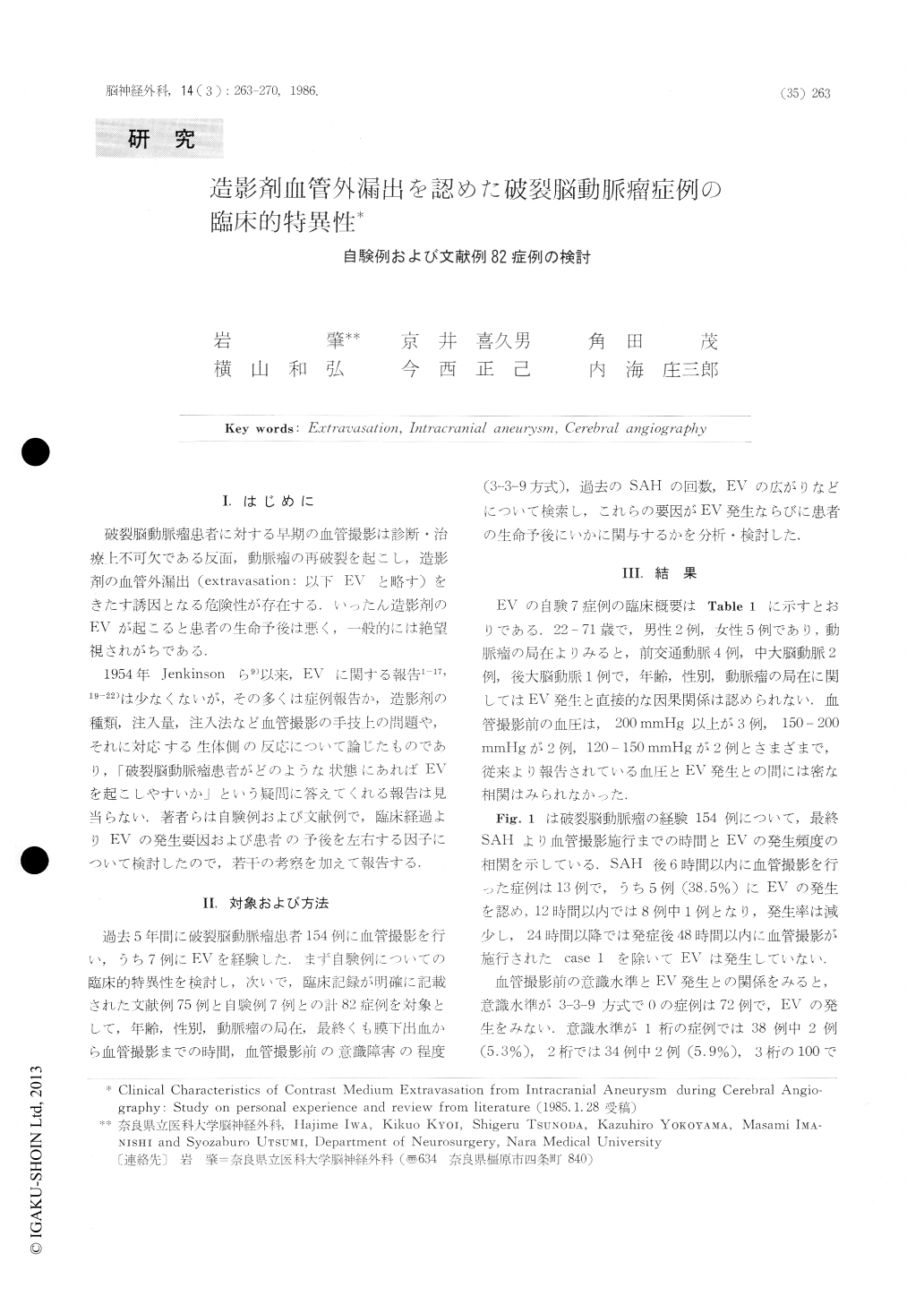
I.はじめに
破裂脳動脈瘤患者に対する早期の血管撮影は診断・治療上不可欠である反面,動脈瘤の再破裂を起こし,造影剤の血管外漏出(extravasation:以下EVと略す)をきたす誘因となる危険性が存在する,いったん造影剤のEVが起こると患者の生命予後は悪く,一般的には絶望視されがちである.
1954年Jenkinsonら9)以来,EVに関する報告1-17,19-22)は少なくないが,その多くは症例報告か,造影剤の種類,注入量,注入法など血管撮影の手技上の問題や,それに対応する生体側の反応について論じたものであり,「破裂脳動脈瘤患者がどのような状態にあればEVを起こしやすいか」という疑問に答えてくれる報告は見当らない.著者らは自験列および文献例で,臨床経過よりEVの発生要因および患者の予後を左右する因子について検討したので,若干の考察を加えて報告する.
Angiography is essential to diagnosis and treatment for the patients with ruptured intracranial aneurysm in early stage, but on the other hand angiography always involves a risk that extravasation (EV) occurs from the aneurysm during angiograhpy. Once EV occurs, the patients outcome is poor and, in general, the patient tends to be regard as hopeless of recovery.
Over the past 5 years, in 154 patients with ruptured intracranial aneurysm angiography was performed, and in 7 of them EV occurred. We performed neck clipping for ruptured aneurysm in 3 of 7 patients and were able to save the life of 2 patients.
Copyright © 1986, Igaku-Shoin Ltd. All rights reserved.