Japanese
English
- 有料閲覧
PPV購入案内
1,320円 (1,200円+税10%) こちらは、875ページから880ページの文献です。購入には医書.jp本会員登録が必要です。
会員登録完了後に改めて上記「購入サイトへ」を選択してください。
または「購入サイトへ」選択後に「購入ログイン」、「新規会員登録」の順に進んでください。
会員登録について詳しくはをご確認ください。
- Abstract 文献概要
- 1ページ目 Look Inside
I.はじめに
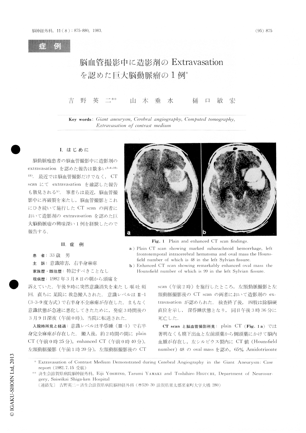
脳動脈瘤患者の脳血管撮影中に造影剤のextravasationを認めた報告は数多い3,4,10,13).最近では脳血管撮影だけでなく,CTscanにてextravasahonを碓認した報告も散見される5).筆者らは最近,脳血管撮影中に再破裂を来たし,脳血管撮影とこれにひき続いて施行したCT scanの両者において造影剤のeMravasationを認めた巨大脳動脈瘤の興味深い1例を経験したので報告する.
The authors report a case of giant aneurysm in which extravasation of contrast medium was demonstrated during cerebral angiography and confirmed by com-puted tomography.
A 33-year-old man suddenly lost consciousness and vomited frequently. Three hours later, he was admitted to our hospital in semicomatose state with left hemiplegia. Within two hours after admission, plain CT scan, enhanced CT scan, left carotid angio-graphy and post-angiographic CT scan were perform-ed. CT scan showed marked subarachnoid hemor-rhage, left temporal intracerebral hematoma and oval mass which was remarkably enhanced in the left Sylvian fissure.
Copyright © 1983, Igaku-Shoin Ltd. All rights reserved.