Japanese
English
- 有料閲覧
PPV購入案内
1,320円 (1,200円+税10%) こちらは、75ページから78ページの文献です。購入には医書.jp本会員登録が必要です。
会員登録完了後に改めて上記「購入サイトへ」を選択してください。
または「購入サイトへ」選択後に「購入ログイン」、「新規会員登録」の順に進んでください。
会員登録について詳しくはをご確認ください。
- Abstract 文献概要
- 1ページ目 Look Inside
Ⅰ.はじめに
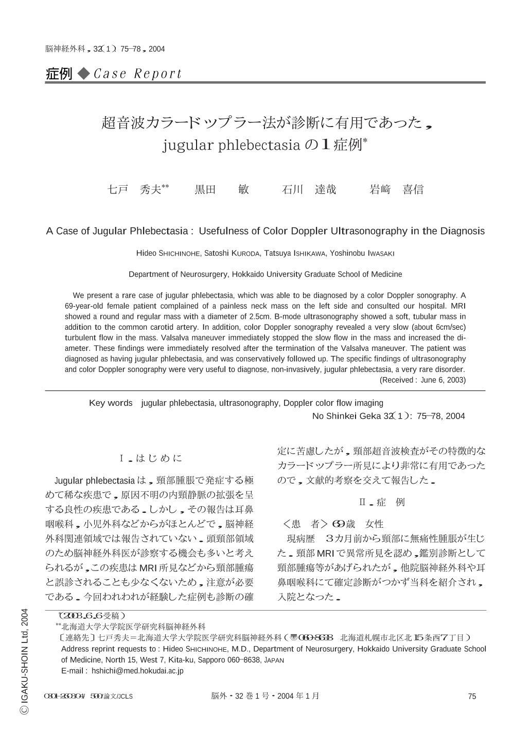
Jugular phlebectasiaは,頸部腫脹で発症する極めて稀な疾患で,原因不明の内頸静脈の拡張を呈する良性の疾患である.しかし,その報告は耳鼻咽喉科,小児外科などからがほとんどで,脳神経外科関連領域では報告されていない.頭頸部領域のため脳神経外科医が診察する機会も多いと考えられるが,この疾患はMRI所見などから頸部腫瘍と誤診されることも少なくないため,注意が必要である.今回われわれが経験した症例も診断の確定に苦慮したが,頸部超音波検査がその特徴的なカラードップラー所見により非常に有用であったので,文献的考察を交えて報告した.
We present a rare case of jugular phlebectasia,which was able to be diagnosed by a color Doppler sonography. A 69-year-old female patient complained of a painless neck mass on the left side and consulted our hospital. MRI showed a round and regular mass with a diameter of 2.5cm. B-mode ultrasonography showed a soft,tubular mass in addition to the common carotid artery. In addition,color Doppler sonography revealed a very slow (about 6cm/sec) turbulent flow in the mass. Valsalva maneuver immediately stopped the slow flow in the mass and increased the diameter. These findings were immediately resolved after the termination of the Valsalva maneuver. The patient was diagnosed as having jugular phlebectasia,and was conservatively followed up. The specific findings of ultrasonography and color Doppler sonography were very useful to diagnose,non-invasively,jugular phlebectasia,a very rare disorder.
Copyright © 2004, Igaku-Shoin Ltd. All rights reserved.