Japanese
English
- 有料閲覧
PPV購入案内
1,320円 (1,200円+税10%) こちらは、79ページから83ページの文献です。購入には医書.jp本会員登録が必要です。
会員登録完了後に改めて上記「購入サイトへ」を選択してください。
または「購入サイトへ」選択後に「購入ログイン」、「新規会員登録」の順に進んでください。
会員登録について詳しくはをご確認ください。
- Abstract 文献概要
- 1ページ目 Look Inside
- 参考文献 Reference
Ⅰ.はじめに
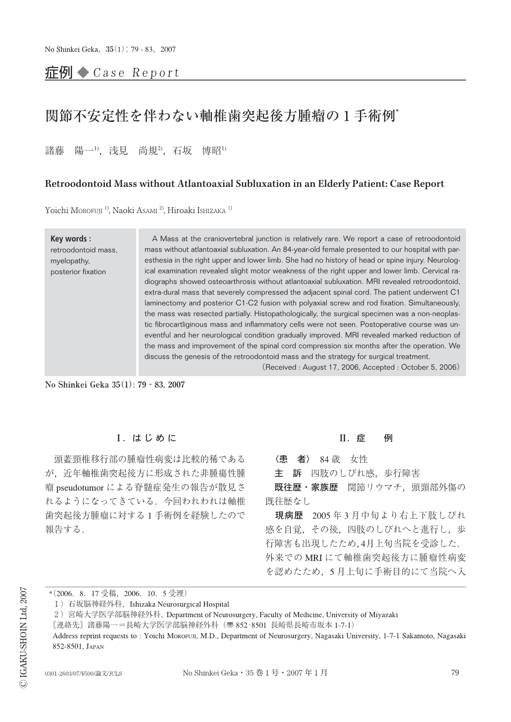
頭蓋頸椎移行部の腫瘤性病変は比較的稀であるが,近年軸椎歯突起後方に形成された非腫瘍性腫瘤pseudotumorによる脊髄症発生の報告が散見されるようになってきている.今回われわれは軸椎歯突起後方腫瘤に対する1手術例を経験したので報告する.
A Mass at the craniovertebral junction is relatively rare. We report a case of retroodontoid mass without atlantoaxial subluxation. An 84-year-old female presented to our hospital with paresthesia in the right upper and lower limb. She had no history of head or spine injury. Neurological examination revealed slight motor weakness of the right upper and lower limb. Cervical radiographs showed osteoarthrosis without atlantoaxial subluxation. MRI revealed retroodontoid, extra-dural mass that severely compressed the adjacent spinal cord. The patient underwent C1 laminectomy and posterior C1-C2 fusion with polyaxial screw and rod fixation. Simultaneously, the mass was resected partially. Histopathologically, the surgical specimen was a non-neoplastic fibrocartliginous mass and inflammatory cells were not seen. Postoperative course was uneventful and her neurological condition gradually improved. MRI revealed marked reduction of the mass and improvement of the spinal cord compression six months after the operation. We discuss the genesis of the retroodontoid mass and the strategy for surgical treatment.
Copyright © 2007, Igaku-Shoin Ltd. All rights reserved.