Japanese
English
- 有料閲覧
PPV購入案内
1,320円 (1,200円+税10%) こちらは、591ページから596ページの文献です。購入には医書.jp本会員登録が必要です。
会員登録完了後に改めて上記「購入サイトへ」を選択してください。
または「購入サイトへ」選択後に「購入ログイン」、「新規会員登録」の順に進んでください。
会員登録について詳しくはをご確認ください。
- Abstract 文献概要
- 1ページ目 Look Inside
- 参考文献 Reference
Ⅰ.は じ め に
脳底部脳主幹脳動脈のアテローム動脈硬化に起因する壁在plaqueは,その成長,破綻により脳梗塞を引き起こす1,2).アテローム血栓性脳梗塞は,虚血性脳血管障害の8~10%を占め2),北米,ヨーロッパ系に比べてアジア系住民でより高い発生率が報告されている10).最近では,薬物治療が無効な症候性狭窄例(>50%)に対して,stentingを含めたballoon angioplastyによる血管内治療を積極的に行うことが推奨されている2).
われわれは,これまで,three-dimensional(3D)MR cisternography(MRC)と3D MR angiography (MRA),3D CT angiography(CTA)とを対比することで,脳底部脳主幹動脈の狭窄・閉塞性病変を評価してきた4).今回,3D MRCと3D MRAとの融合画像(3D MRC/MRA fusion image)を作成し6,7,9),アテローム動脈硬化による頭蓋内脳主幹動脈の狭窄性病変を血管外壁形態とともに立体的に画像評価したので,fusion imagingによる画像解析技術を報告し,その利点と問題点につき検討した.
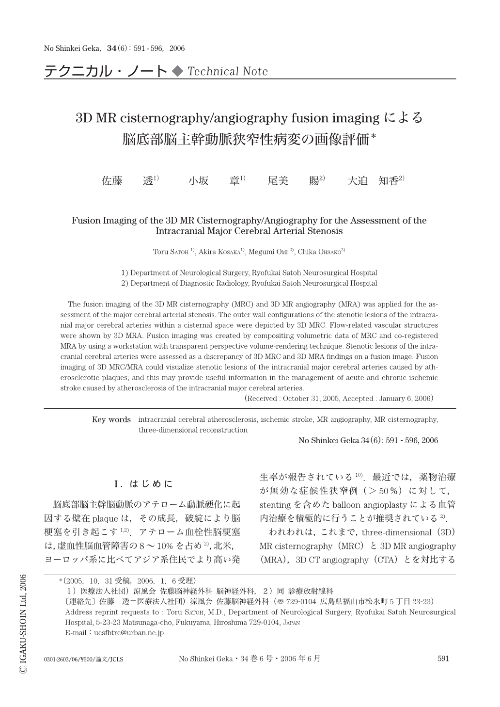
The fusion imaging of the 3D MR cisternography (MRC) and 3D MR angiography (MRA) was applied for the assessment of the major cerebral arterial stenosis. The outer wall configurations of the stenotic lesions of the intracranial major cerebral arteries within a cisternal space were depicted by 3D MRC. Flow-related vascular structures were shown by 3D MRA. Fusion imaging was created by compositing volumetric data of MRC and co-registered MRA by using a workstation with transparent perspective volume-rendering technique. Stenotic lesions of the intracranial cerebral arteries were assessed as a discrepancy of 3D MRC and 3D MRA findings on a fusion image. Fusion imaging of 3D MRC/MRA could visualize stenotic lesions of the intracranial major cerebral arteries caused by atherosclerotic plaques; and this may provide useful information in the management of acute and chronic ischemic stroke caused by atherosclerosis of the intracranial major cerebral arteries.
Copyright © 2006, Igaku-Shoin Ltd. All rights reserved.