Japanese
English
- 有料閲覧
- Abstract 文献概要
- 1ページ目 Look Inside
Ⅰ.はじめに
脳動脈瘤の発生,成長,破裂に至るメカニズムは,これまで脳動脈瘤の大きさやbleb形成などの形態学的要素1,27,28),脳動脈瘤内の血流パターンやshear stressなどの流体力学的要素3,9,22,24,26),脳動脈瘤壁の弾力性や脆弱性などの病理学的要素6,8)など,主として脳動脈瘤血管構築および瘤内環境について報告されてきた.近年,脳動脈瘤外壁とこれに接触,癒着する脳実質や脳神経など脳動脈瘤周囲構造物との解剖学的関係が,脳動脈瘤の瘤外・瘤周囲環境(perianeurymal environment)として臨床的に検討され,特に脳動脈瘤の破裂や脳神経症状の発症に関わる要因の1つとして関心がもたれる14,17).
T2-weighted three-dimensional(3D)fast spin-echo(FSE)sequence(heavy T2-weighted imaging)などで得られるMR脳槽画像(MR cisternogram,MRC)では,信号雑音比が良好で微細な断層情報(volume data)が得られ,脳槽内脳脊髄液は高信号強度で,脳動脈瘤や親動脈,脳表静脈・架橋静脈,脳神経などの脳槽内構造物や脳実質,硬膜・頭蓋底骨構造は低信号強度で描出される7,10-13,15,16).これにより,MRCでは,脳動脈瘤の外壁は脳脊髄液と明瞭に境界され,脳槽内陰影欠損像として,周囲脳実質,脳表静脈,脳神経構造物などの瘤外・瘤周囲環境とともに表示可能となる.
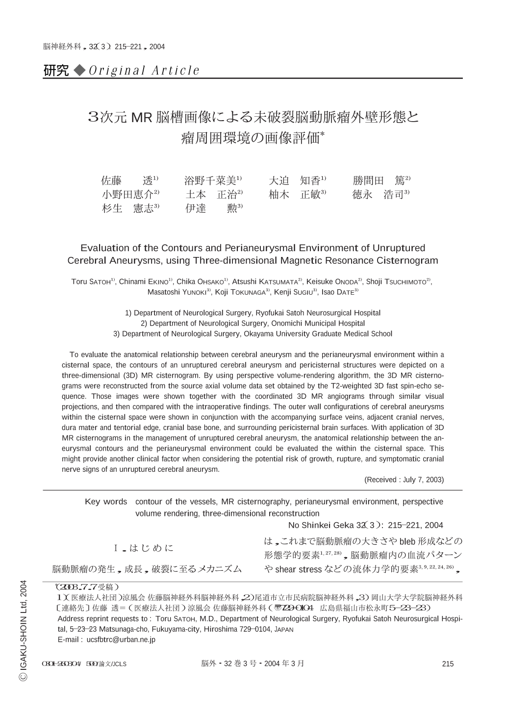
To evaluate the anatomical relationship between cerebral aneurysm and the perianeurysmal environment within a cisternal space,the contours of an unruptured cerebral aneurysm and pericisternal structures were depicted on a three-dimensional (3D) MR cisternogram. By using perspective volume-rendering algorithm,the 3D MR cisternograms were reconstructed from the source axial volume data set obtained by the T2-weighted 3D fast spin-echo sequence. Those images were shown together with the coordinated 3D MR angiograms through similar visual projections,and then compared with the intraoperative findings. The outer wall configurations of cerebral aneurysms within the cisternal space were shown in conjunction with the accompanying surface veins,adjacent cranial nerves,dura mater and tentorial edge,cranial base bone,and surrounding pericisternal brain surfaces. With application of 3D MR cisternograms in the management of unruptured cerebral aneurysm,the anatomical relationship between the aneurysmal contours and the perianeurysmal environment could be evaluated the within the cisternal space. This might provide another clinical factor when considering the potential risk of growth,rupture,and symptomatic cranial nerve signs of an unruptured cerebral aneurysm.

Copyright © 2004, Igaku-Shoin Ltd. All rights reserved.


