Japanese
English
- 有料閲覧
- Abstract 文献概要
- 1ページ目 Look Inside
- 参考文献 Reference
Ⅰ.はじめに
脳動脈瘤の発生,成長,破裂のメカニズムは,これまで親動脈と脳動脈瘤との血管構築,瘤サイズと形態,瘤壁性状,瘤内血行動態など主として脳動脈瘤自体が有する瘤内環境について検討されてきた1-4,13,16,18-22).これらに加え,脳動脈瘤を取り囲む瘤周囲環境(perianeurysmal environment)は,脳動脈瘤の伸展やdomeの変形にかかわる瘤外要因と考えられている10,15,17).
内頸動脈-後交通動脈瘤は成長・増大に伴い,硬膜・硬膜襞,天幕自由縁,後床突起,動眼神経,後交通動脈,さらには側頭葉内側面など多くの瘤周囲構造物と接触する可能性を有する.今回われわれは,未破裂内頸動脈-後交通動脈瘤において,MR cisternography (MRC)とMR angiography (MRA)とを連続して撮像し,得られたvolume dataからthree-dimensional (3D) MRCとその等座標3D MRAを画像再構成した.さらに,両者のvolume dataを重畳した3D MRC-MRA fusion imageを新たに創作し,未破裂脳動脈瘤の伸展・変形に及ぼす瘤周囲環境に着目して画像解析したので報告する.
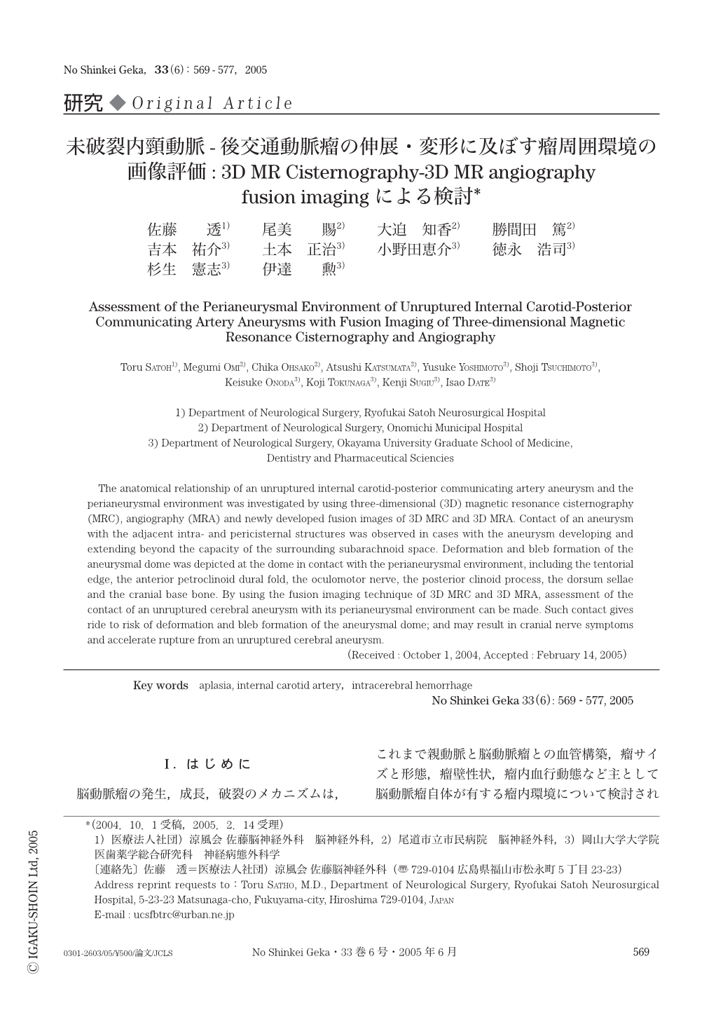
The anatomical relationship of an unruptured internal carotid-posterior communicating artery aneurysm and the perianeurysmal environment was investigated by using three-dimensional (3D) magnetic resonance cisternography (MRC), angiography (MRA) and newly developed fusion images of 3D MRC and 3D MRA. Contact of an aneurysm with the adjacent intra- and pericisternal structures was observed in cases with the aneurysm developing and extending beyond the capacity of the surrounding subarachnoid space. Deformation and bleb formation of the aneurysmal dome was depicted at the dome in contact with the perianeurysmal environment, including the tentorial edge, the anterior petroclinoid dural fold, the oculomotor nerve,the posterior clinoid process, the dorsum sellae and the cranial base bone. By using the fusion imaging technique of 3D MRC and 3D MRA,assessment of the contact of an unruptured cerebral aneurysm with its perianeurysmal environment can be made. Such contact gives ride to risk of deformation and bleb formation of the aneurysmal dome; and may result in cranial nerve symptoms and accelerate rupture from an unruptured cerebral aneurysm.

Copyright © 2005, Igaku-Shoin Ltd. All rights reserved.


