Japanese
English
- 有料閲覧
PPV購入案内
1,320円 (1,200円+税10%) こちらは、441ページから451ページの文献です。購入には医書.jp本会員登録が必要です。
会員登録完了後に改めて上記「購入サイトへ」を選択してください。
または「購入サイトへ」選択後に「購入ログイン」、「新規会員登録」の順に進んでください。
会員登録について詳しくはをご確認ください。
- Abstract 文献概要
- 1ページ目 Look Inside
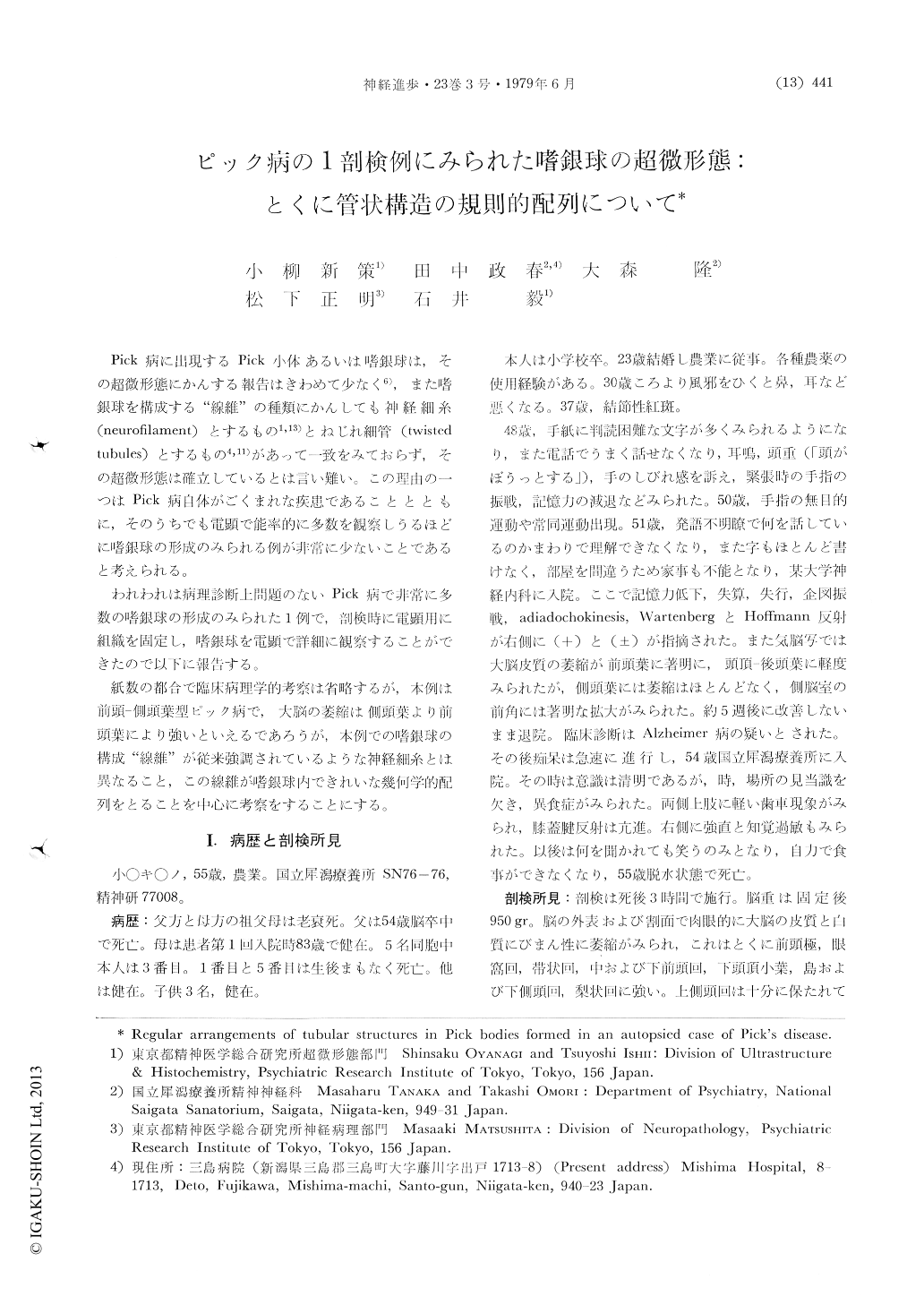
Pick病に出現するPick小体あるいは嗜銀球は,その超微形態にかんする報告はきわめて少なく6),また嗜銀球を構成する"線維"の種類にかんしても神経細糸(neurofilament)とするもの1,13)とねじれ細管(twistedtubules)とするもの4,11)があって一致をみておらず,その超微形態は確立しているとは言い難い。この理由の一つはPick病自体がごくまれな疾患であることとともに,そのうちでも電顕で能率的に多数を観察しうるほどに嗜銀球の形成のみられる例が非常に少ないことであると考えられる。
われわれは病理診断上問題のないPick病で非常に多数の嗜銀球の形成のみられた1例で,剖検時に電顕用に組織を固定し,嗜銀球を電顕で詳細に観察することができたので以下に報告する。
Abstract
Tissue for examination by the electron micro-scope was taken out at autopsy 3 hours afterdeath from the hippocampus, dentate gyrus, inferior temporal gyrus and superior frontal gyrus of a case of Pick's disease, 55 years old, female, with a duration of 7 years.
Brain weighed 950 gm. Pathologically, cortical atrophy and subcortical gliosis were particularly prominent in middle and inferior frontal gyrus, orbital gyrus, rectal gyrus, inferior parietal lobule, insula, middle and inferior temporal gyrus and fusiform gyrus (Fig. 1 & 2).
Copyright © 1979, Igaku-Shoin Ltd. All rights reserved.