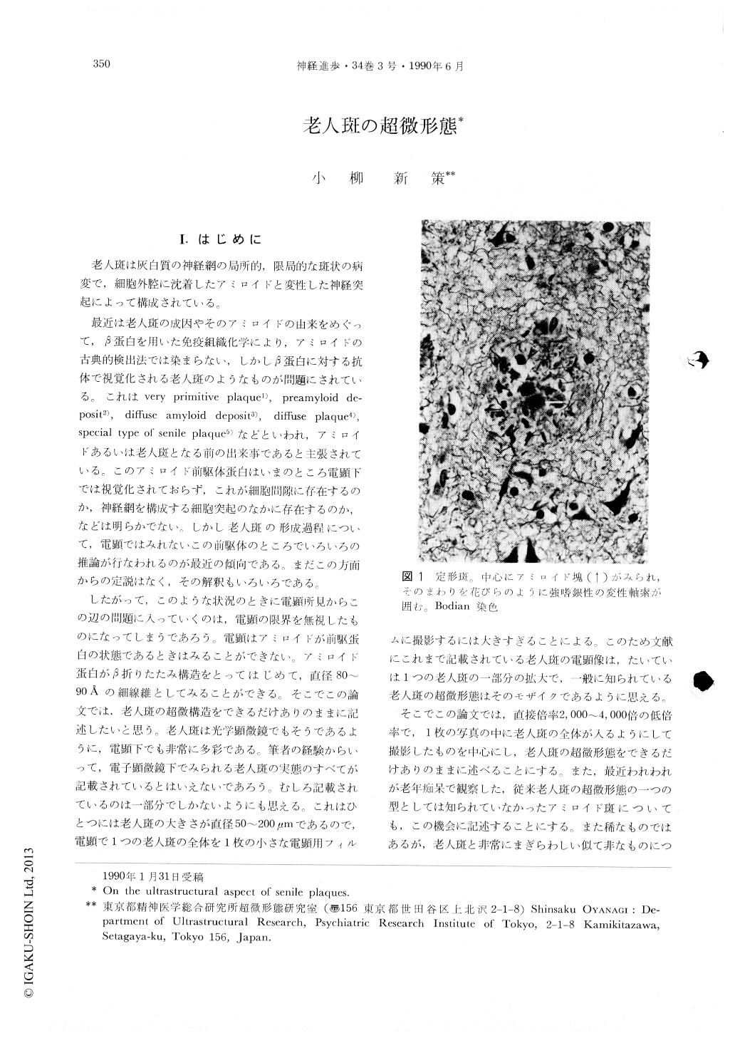
Japanese
English
- 有料閲覧
- Abstract 文献概要
- 1ページ目 Look Inside
I.はじめに
老人斑は灰白質の神経網の局所的,限局的な斑状の病変で,細胞外腔に沈着したアミロイドと変性した神経突起によって構成されている。
最近は老人斑の成因やそのアミロイドの由来をめぐって,β蛋白を用いた免疫組織化学により,アミロイドの古典的検出法では染まらない,しかしβ蛋白に対する抗体で視覚化される老人斑のようなものが問題にされている。これはvery primitive plaque1),preamyloid deposit2),diffuse amyloid deposit3),diffuse plaque4),special type of senile plaque5)などといわれ,アミロイドあるいは老人斑となる前の出来事であると主張されている。このアミロイド前駆体蛋白はいまのところ電顕下では視覚化されておらず,これが細胞間隙に存在するのか,神経網を構成する細胞突起のなかに存在するのか,などは明らかでない。しかし老人斑の形成過程について,電顕ではみれないこの前駆体のところでいろいろの推論が行なわれるのが最近の傾向である。まだこの方面からの定説はなく,その解釈もいろいろである。
Several reports and reviews describing ultrastructure of senile plaques appeared particularly in the years between 1964 and 1975, however, such ultrastructural studies have been slowing down since that time. Wisniewski and Terry categorized senile plaques into three types, that is, primitive plaque, typical plaque and core plaque on the basis of their ultrastructural observation in 1973. It is obvious, however, that the ultrastructural aspect of senile plaques has not fully been described, because appearance of senile plaques is varied under the electron microscope and the size of the senile plaque is so large, 50-200 mμ in diameter, that it has been difficult to take photographs embodying the entire feature of a senile plaque on a sheet of the film.

Copyright © 1990, Igaku-Shoin Ltd. All rights reserved.


