Japanese
English
- 有料閲覧
PPV購入案内
1,320円 (1,200円+税10%) こちらは、902ページから908ページの文献です。購入には医書.jp本会員登録が必要です。
会員登録完了後に改めて上記「購入サイトへ」を選択してください。
または「購入サイトへ」選択後に「購入ログイン」、「新規会員登録」の順に進んでください。
会員登録について詳しくはをご確認ください。
- Abstract 文献概要
- 1ページ目 Look Inside
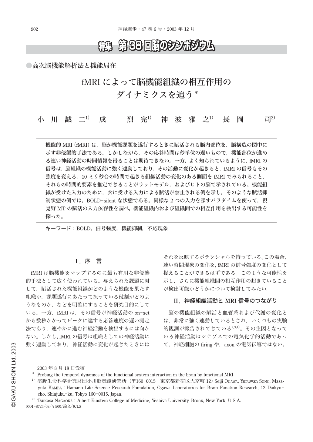
機能的MRI(fMRI)は,脳が機能課題を遂行するときに賦活される脳内部位を,脳構造の図中に示す非侵襲的手法である。しかしながら,その応答時間は秒単位の遅いもので,機能部位が進める速い神経活動の時間情報を得ることは期待できない。一方,よく知られているように,fMRIの信号は,脳組織の機能活動に強く連動しており,その活動に変化が起きると,fMRIの信号もその強度を変える。10ミリ秒台の時間で起きる組織活動の変化のある側面をfMRIでみられること,それらの時間的要素を推定できることがラットモデル,およびヒトの脳で示されている。機能組織が受けた入力のために,次に受ける入力による賦活が禁止される例を示し,そのような賦活抑制状態の例では,BOLD-silentな状態である。同様な2つの入力を課すパラダイムを使って,視覚野MTの賦活の入力依存性を調べ,機能組織内および組織間での相互作用を検出する可能性を探った。
fMRI is known to be a method to find locations of sites in the brain which respond to a given functional task. The response time of fMRI signal, however, is at the order of seconds and too slow to detect any temporally fast neural process a functional system takes to perform a task. It is also well-known that fMRI signal is tightly coupled to such neural process and sensitive to show a change in signal amplitude when the neural system-activity changes. In rat models as well as in the human brain, we show that such signal changes responding to activity variation are observable even it occurs within 10's milliseconds or hundreds milliseconds. Using pair wise stimulation paradigms, we have examined these signal responses in MT to characterize some aspects of a neuro-refractory response. We pursue the potential of such approach to detect interaction among sites activated by performing some functional task.
Copyright © 2003, Igaku-Shoin Ltd. All rights reserved.