Japanese
English
- 有料閲覧
PPV購入案内
1,320円 (1,200円+税10%) こちらは、962ページから967ページの文献です。購入には医書.jp本会員登録が必要です。
会員登録完了後に改めて上記「購入サイトへ」を選択してください。
または「購入サイトへ」選択後に「購入ログイン」、「新規会員登録」の順に進んでください。
会員登録について詳しくはをご確認ください。
- Abstract 文献概要
- 1ページ目 Look Inside
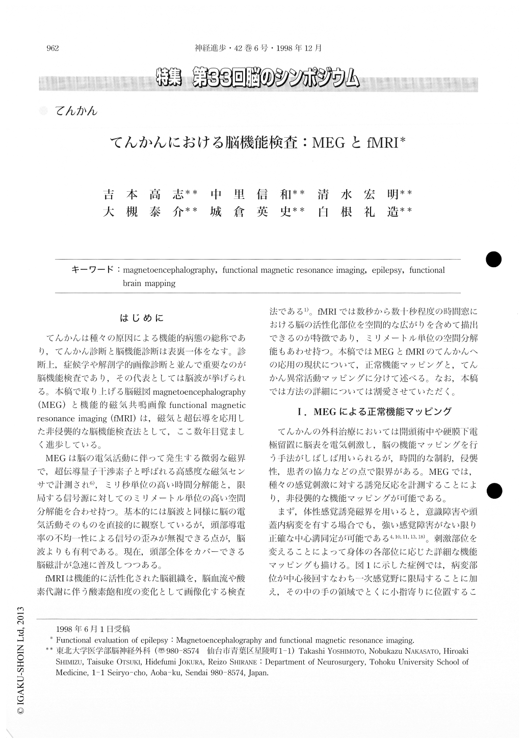
はじめに
てんかんは種々の原因による機能的病態の総称であり,てんかん診断と脳機能診断は表裏一体をなす。診断上,症候学や解剖学的画像診断と並んで重要なのが脳機能検査であり,その代表としては脳波が挙げられる。本稿で取り上げる脳磁図magnetoencephalography(MEG)と機能的磁気共嶋画像functional magneticresonance imaging(fMRI)は,磁気と超伝導を応用した非侵襲的な脳機能検査法として,ここ数年目覚ましく進歩している。
MEGは脳の電気活動に伴って発生する微弱な磁界で,超伝導量子干渉素子と呼ばれる高感度な磁気センサで計測され6),ミリ秒単位の高い時間分解能と,限局する信号源に対してのミリメートル単位の高い空間分解能を合わせ持つ。基本的には脳波と同様に脳の電気活動そのものを直接的に観察しているが,頭部導電率の不均一性による信号の歪みが無視できる点が,脳波よりも有利である。現在,頭部全体をカバーできる脳磁計が急速に普及しつつある。
The clinical utility of magnetoencephalography (MEG) and functional magnetic resonance imaging (fMRI) is reviewed for evaluating both normal and abnormal brain function in patients with intractable epilepsy. Recent advances in magnetophysiology and superconductivity physics have enabled non-invasive multimodal functional brain imaging. MEG can be applied to mapping of the somatosensory, auditory and visual cortices. The sources of the evoked magnetic fields are localized near the primary sensory area. Latency delay in the evoked responses can be used for quantitative evaluation of brain function.
Copyright © 1998, Igaku-Shoin Ltd. All rights reserved.