Japanese
English
- 有料閲覧
PPV購入案内
1,320円 (1,200円+税10%) こちらは、949ページから953ページの文献です。購入には医書.jp本会員登録が必要です。
会員登録完了後に改めて上記「購入サイトへ」を選択してください。
または「購入サイトへ」選択後に「購入ログイン」、「新規会員登録」の順に進んでください。
会員登録について詳しくはをご確認ください。
- Abstract 文献概要
- 1ページ目 Look Inside
- 参考文献 Reference
はじめに
今回われわれは,慢性肉芽腫性疾患,特に神経サルコイドーシスと脳腫瘍との鑑別が困難であった胚芽腫の1例を経験したので報告する。
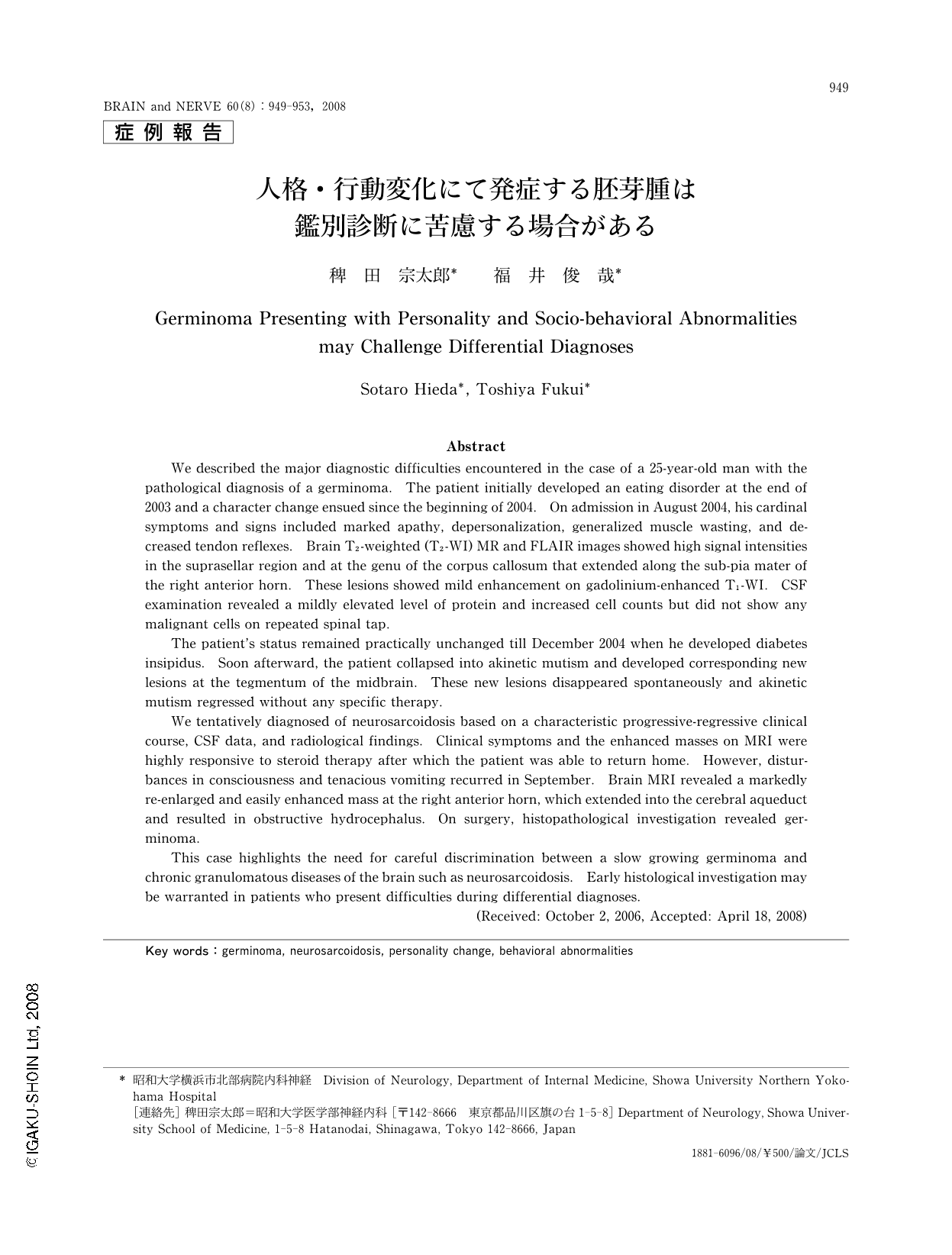
Abstract
We described the major diagnostic difficulties encountered in the case of a 25-year-old man with the pathological diagnosis of a germinoma. The patient initially developed an eating disorder at the end of 2003 and a character change ensued since the beginning of 2004. On admission in August 2004, his cardinal symptoms and signs included marked apathy, depersonalization, generalized muscle wasting, and decreased tendon reflexes. Brain T2-weighted (T2-WI) MR and FLAIR images showed high signal intensities in the suprasellar region and at the genu of the corpus callosum that extended along the sub-pia mater of the right anterior horn. These lesions showed mild enhancement on gadolinium-enhanced T1-WI. CSF examination revealed a mildly elevated level of protein and increased cell counts but did not show any malignant cells on repeated spinal tap.
The patient's status remained practically unchanged till December 2004 when he developed diabetes insipidus. Soon afterward, the patient collapsed into akinetic mutism and developed corresponding new lesions at the tegmentum of the midbrain. These new lesions disappeared spontaneously and akinetic mutism regressed without any specific therapy.
We tentatively diagnosed of neurosarcoidosis based on a characteristic progressive-regressive clinical course, CSF data, and radiological findings. Clinical symptoms and the enhanced masses on MRI were highly responsive to steroid therapy after which the patient was able to return home. However, disturbances in consciousness and tenacious vomiting recurred in September. Brain MRI revealed a markedly re-enlarged and easily enhanced mass at the right anterior horn, which extended into the cerebral aqueduct and resulted in obstructive hydrocephalus. On surgery, histopathological investigation revealed germinoma.
This case highlights the need for careful discrimination between a slow growing germinoma and chronic granulomatous diseases of the brain such as neurosarcoidosis. Early histological investigation may be warranted in patients who present difficulties during differential diagnoses.
Copyright © 2008, Igaku-Shoin Ltd. All rights reserved.