Japanese
English
- 有料閲覧
PPV購入案内
1,320円 (1,200円+税10%) こちらは、859ページから865ページの文献です。購入には医書.jp本会員登録が必要です。
会員登録完了後に改めて上記「購入サイトへ」を選択してください。
または「購入サイトへ」選択後に「購入ログイン」、「新規会員登録」の順に進んでください。
会員登録について詳しくはをご確認ください。
- Abstract 文献概要
- 1ページ目 Look Inside
- 参考文献 Reference
Ⅰ.はじめに
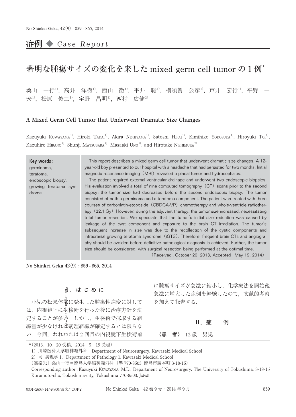
小児の松果体部に発生した腫瘍性病変に対しては,内視鏡下に生検術を行った後に治療方針を決定することが多い.しかし,生検術で採取する組織量が少なければ病理組織が確定するとは限らない.今回,われわれは2回目の内視鏡下生検術前に腫瘍サイズが急激に縮小し,化学療法を開始後急激に増大した症例を経験したので,文献的考察を加えて報告する.
This report describes a mixed germ cell tumor that underwent dramatic size changes. A 12-year-old boy presented to our hospital with a headache that had persisted for two months. Initial magnetic resonance imaging(MRI)revealed a pineal tumor and hydrocephalus.
The patient required external ventricular drainage and underwent two endoscopic biopsies. His evaluation involved a total of nine computed tomography(CT)scans prior to the second biopsy;the tumor size had decreased before the second endoscopic biopsy. The tumor consisted of both a germinoma and a teratoma component. The patient was treated with three courses of carboplatin-etoposide(CBDCA-VP)chemotherapy and whole-ventricle radiotherapy(32.1Gy). However, during the adjuvant therapy, the tumor size increased, necessitating total tumor resection. We speculate that the tumor’s initial size reduction was caused by leakage of the cyst component and exposure to the brain CT irradiation. The tumor’s subsequent increase in size was due to the recollection of the cystic components and intracranial growing teratoma syndrome(iGTS). Therefore, frequent brain CTs and angiography should be avoided before definitive pathological diagnosis is achieved. Further, the tumor size should be considered, with surgical resection being performed at the optimal time.
Copyright © 2014, Igaku-Shoin Ltd. All rights reserved.