Japanese
English
- 有料閲覧
PPV購入案内
1,320円 (1,200円+税10%) こちらは、431ページから433ページの文献です。購入には医書.jp本会員登録が必要です。
会員登録完了後に改めて上記「購入サイトへ」を選択してください。
または「購入サイトへ」選択後に「購入ログイン」、「新規会員登録」の順に進んでください。
会員登録について詳しくはをご確認ください。
- Abstract 文献概要
- 1ページ目 Look Inside
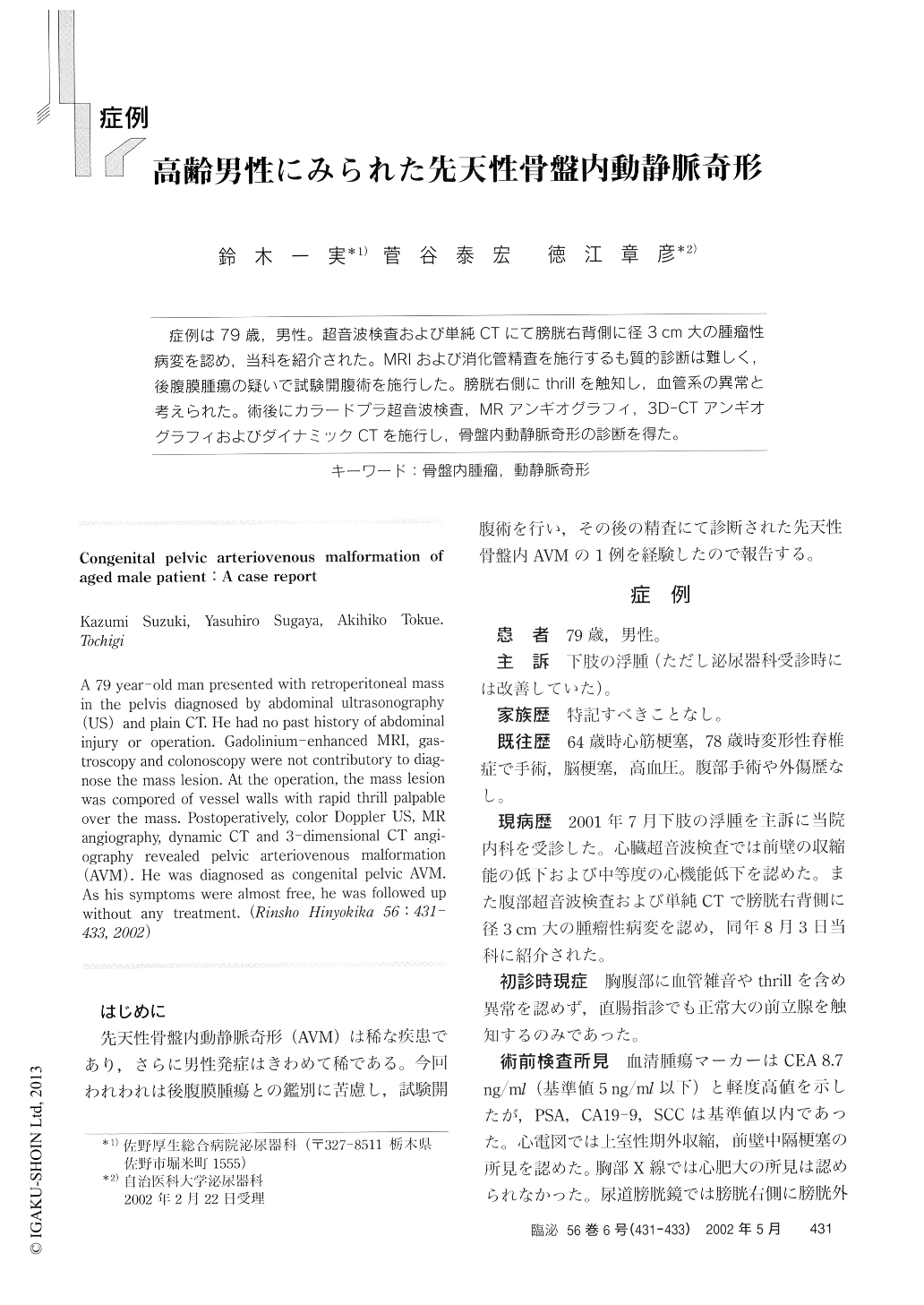
症例は79歳,男性。超音波検査および単純CTにて膀胱右背側に径3cm大の腫瘤性病変を認め,当科を紹介された。MRIおよび消化管精査を施行するも質的診断は難しく,後腹膜腫瘍の疑いで試験開腹術を施行した。膀胱右側にthrillを触知し,血管系の異常と考えられた。術後にカラードプラ超音波検査,MRアンギオグラフィ,3D-CTアンギオグラフィおよびダイナミックCTを施行し,骨盤内動静脈奇形の診断を得た。
A 79 year-old man presented with retroperitoneal mass in the pelvis diagnosed by abdominal ultrasonography (US) and plain CT. He had no past history of abdominal injury or operation. Gadolinium-enhanced MRI, gas-troscopy and colonoscopy were not contributory to diag-nose the mass lesion. At the operation, the mass lesion was compored of vessel walls with rapid thrill palpable over the mass. Postoperatively, color Doppler US, MR angiography, dynamic CT and 3-dimensional CT angi-ography revealed pelvic arteriovenous malformation (AVM). He was diagnosed as congenital pelvic AVM.
Copyright © 2002, Igaku-Shoin Ltd. All rights reserved.