Japanese
English
- 有料閲覧
PPV購入案内
1,320円 (1,200円+税10%) こちらは、269ページから272ページの文献です。購入には医書.jp本会員登録が必要です。
会員登録完了後に改めて上記「購入サイトへ」を選択してください。
または「購入サイトへ」選択後に「購入ログイン」、「新規会員登録」の順に進んでください。
会員登録について詳しくはをご確認ください。
- Abstract 文献概要
- 1ページ目 Look Inside
緒言
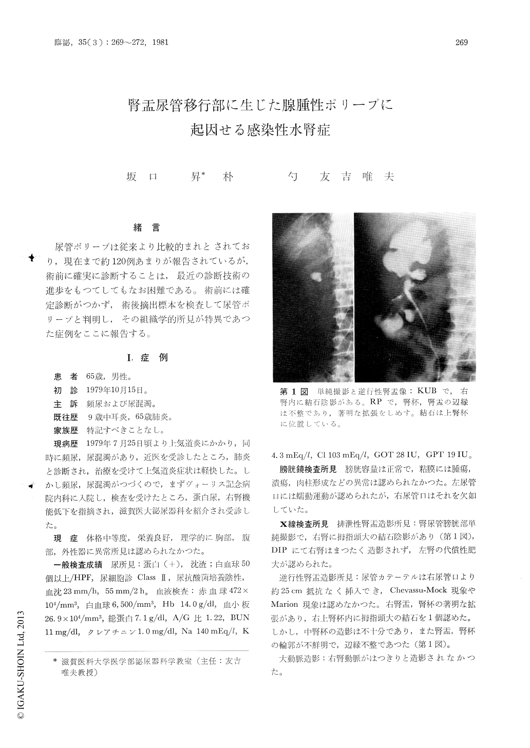
尿管ポリープは従来より比較的まれとされており,現在まで約120例あまりが報告されているが,術前に確実に診断することは,最近の診断技術の進歩をもつてしてもなお困難である。術前には確定診断がつかず,術後摘出標本を検査して尿管ポリープと判明し,その組織学的所見が特異であつた症例をここに報告する。
A 65-year-old man was admitted with intractable urinary tract infection. KUB showed a stone in the right kidney. IVP revealed right nonvisualized kidney, which had no uptake of radionuclide on renal scintigraphy and extremely poor vascularity on angiography. Retrograde pyelography suggested infected hydronephrosis with a calculus. Right nephrectomy was performed. The removed kidney, weighing 150 grams, showed the feature of infected hydronephrosis. At the ureteropelvic junction, were found two polyps each measuring 1.5cm×0.7cm. A pathohistological study showed chronic pyelone-phritis with contracted parenchyma and adcnomatous polyps.
Copyright © 1981, Igaku-Shoin Ltd. All rights reserved.