Japanese
English
- 有料閲覧
PPV購入案内
1,320円 (1,200円+税10%) こちらは、17ページから23ページの文献です。購入には医書.jp本会員登録が必要です。
会員登録完了後に改めて上記「購入サイトへ」を選択してください。
または「購入サイトへ」選択後に「購入ログイン」、「新規会員登録」の順に進んでください。
会員登録について詳しくはをご確認ください。
- Abstract 文献概要
- 1ページ目 Look Inside
Ⅰ.はじめに
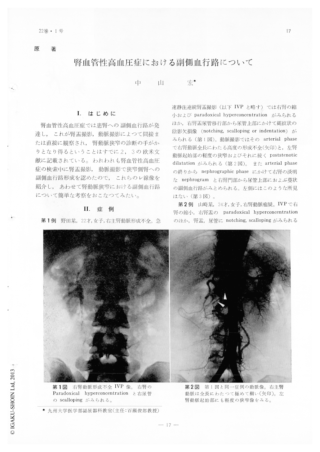
腎血管性高血圧症では患腎への副側血行路が発達し,これが腎盂撮影,動脈撮影によつて間接または直接に観察され,腎動脈狭窄の診断の手がかりとなり得るということはすでに2,3の欧米文献に記載されている。われわれも腎血管性高血圧症の検索中に腎盂撮影,動脈撮影で狭窄側腎への副側血行路形成を認めたので,これらのレ線像を紹介し,あわせて腎動脈狭窄における副側血行路について簡単な考察をおこなつてみたい。
The demonstration of collateral pathways to an ischemic kidney seems to be of great value in the diagnosis of renovascular hypertension. Collateral vessels which develop around the upper urinary tract can be visualized indirectly in a urogram as a characteristic sign of"notching"or"scalloping"of the renal pelvis and/or the ureter.
Four, among the 5 cases of renovascular hypertension reported here, showed marked development of collateral vessels both in urogram and in angiogram, in one case, however, no collateral circulation could be demonstrated in spite of the presence of the right renal artery stenosis.
Copyright © 1968, Igaku-Shoin Ltd. All rights reserved.