Japanese
English
- 有料閲覧
PPV購入案内
1,320円 (1,200円+税10%) こちらは、629ページから639ページの文献です。購入には医書.jp本会員登録が必要です。
会員登録完了後に改めて上記「購入サイトへ」を選択してください。
または「購入サイトへ」選択後に「購入ログイン」、「新規会員登録」の順に進んでください。
会員登録について詳しくはをご確認ください。
- Abstract 文献概要
- 1ページ目 Look Inside
I.はじめに
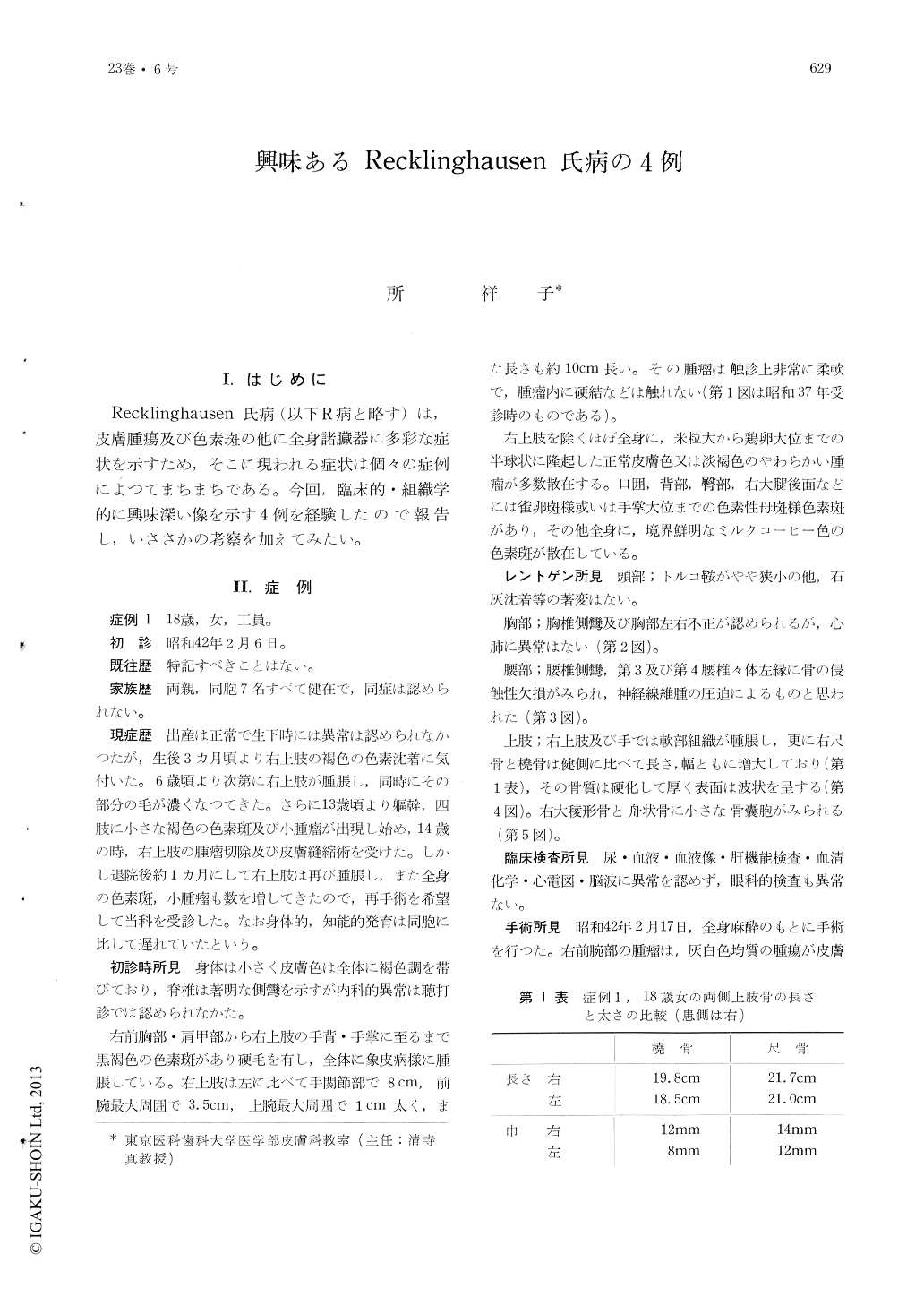
Recklinghausen氏病(以下R病と略す)は,皮膚腫瘍及び色素斑の他に全身諸臓器に多彩な症状を示すため,そこに現われる症状は個々の症例によつてまちまちである。今回,臨床的・組織学的に興味深い像を示す4例を経験したので報告し,いささかの考察を加えてみたい。
Four cases of this disease were reported.
Case 1: An 18-year-old female with typical pigmented spots and disseminated skin tumors had a large tumor which covered the entire right, upper extremity. Its roentgenogram revealed abnormal development of the brachial, radial and ulnar bones, scoliosis, erosive defect and cyst formation of bone. Its histologic picture was so-called neurofibroma.
Case 2: A 17-year-old female had pigmented macules, skin tumors, subcutaneous nodules arranged in rosary and scoliosis. The subcutaneous nodules were composed of myelinated nerve fibers, most of which showed slight degeneration.
Case 3 : A 25-year-old, elder brother of case 2, had pigmented spots and skin tumors.
Case 4 : A 6-year-old girl showed pigmented spots which were disseminated over the entire body surface, and large pigmented hairy spots in the gluteal and right femoral regions under which soft tumors were found. She also had spina bifida and erosive defect of the pelvic bones. Histologic specimen from the skin tumor was composed partly of a so-called neurofibromatous structure and partly of severely degenerated neuromatous structure.
Pathologic changes of nerve fibers such as those seen in case 2 might be an early picture of this disease, which might gradually degenerate into such a situation as seen in case 4.
Relationships between proliferation of nerve fibers and that of schwannian cells were not de-monstrated.
Copyright © 1969, Igaku-Shoin Ltd. All rights reserved.