Japanese
English
- 有料閲覧
PPV購入案内
1,320円 (1,200円+税10%) こちらは、66ページから69ページの文献です。購入には医書.jp本会員登録が必要です。
会員登録完了後に改めて上記「購入サイトへ」を選択してください。
または「購入サイトへ」選択後に「購入ログイン」、「新規会員登録」の順に進んでください。
会員登録について詳しくはをご確認ください。
- Abstract 文献概要
- 1ページ目 Look Inside
はじめに
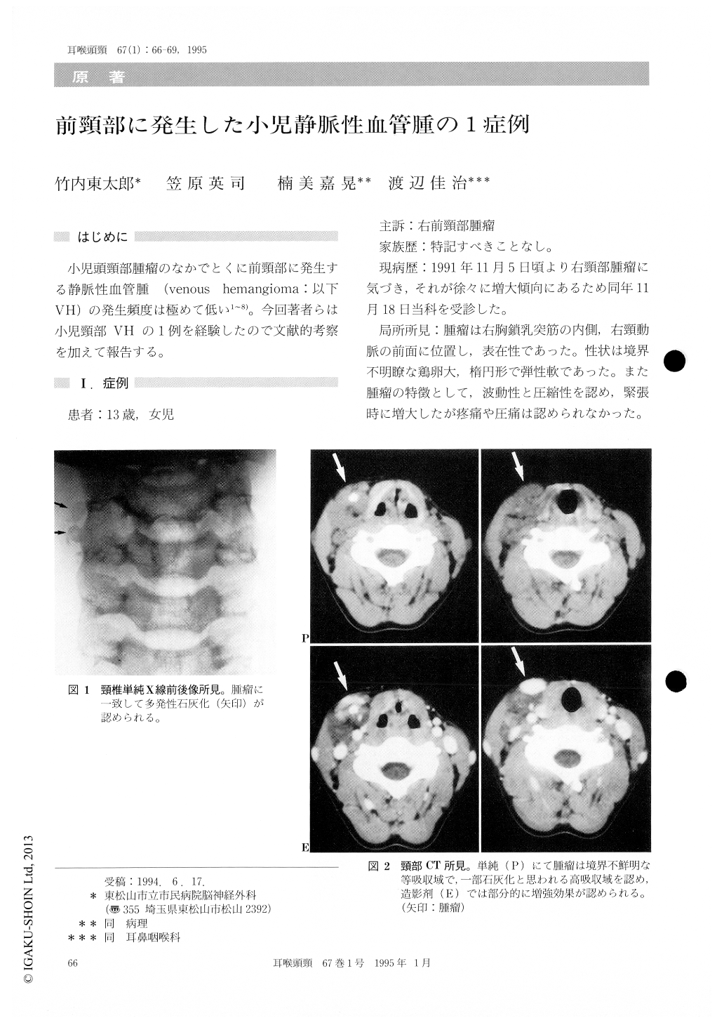
小児頭頸部腫瘤のなかでとくに前頸部に発生する静脈性血管腫(venous hemangioma:以下VH)の発生頻度は極めて低い1〜8)。今回著者らは小児頸部VHの1例を経験したので文献的考察を加えて報告する。
A 13-year-old girl presented with a mass in the right cervical region. Since it was gradually enlarg-ing, she visited our department. The mass was hen's egg-sized and ovoid, and located in the medial side of the right sternomastoid muscle. It was found to be compressed, and was enlarged during tension. CT of the neck revealed isodensity and high density, part of which seemed to indicate calcification. Partial enhancement with contrast medium was observed. T2-weighted image of MRI revealed high signal intensity. Angiogram of the neck revealed a shadow of abnormal blood vessel centering of the venous phase. Excision was performed for cosmetic purposes. The tumor was found associated with nutritional blood vessels from the external carotid artery and communicated with the right jugular vein. Histopathological study showed venous hemangioma.
Copyright © 1995, Igaku-Shoin Ltd. All rights reserved.