Japanese
English
- 有料閲覧
PPV購入案内
1,320円 (1,200円+税10%) こちらは、933ページから936ページの文献です。購入には医書.jp本会員登録が必要です。
会員登録完了後に改めて上記「購入サイトへ」を選択してください。
または「購入サイトへ」選択後に「購入ログイン」、「新規会員登録」の順に進んでください。
会員登録について詳しくはをご確認ください。
- Abstract 文献概要
- 1ページ目 Look Inside
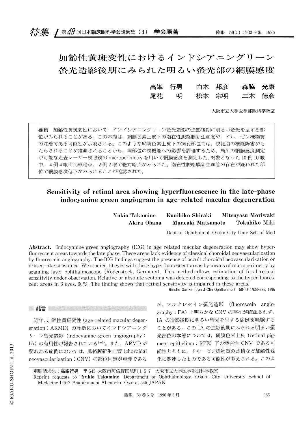
加齢性黄斑変性において,インドシアニングリーン螢光造影の造影後期に明るい螢光を呈する部位がみられることがある。この本態は,網膜色素上皮下の潜在性脈絡膜新生血管や,ドルーゼン様物質の沈着である可能性が示唆される。このような網膜色素上皮下の病変部位では,視細胞の機能障害がもたらされることが推測されることから,同部位の視機能への影響を評価するため,局所の網膜感度測定が可能な走査レーザー検眼鏡のmicroperimetryを用いて網膜感度を測定した。対象となった10例10眼中,4例4眼で比較暗点,2例2眼で絶対暗点がみられた。潜在性脈絡膜新生血管の存在が疑われた部位で網膜感度低下がみられることが確認された。
Indocyanine green angiography (ICG) in age-related macular degeneration may show hyper-fluorescent areas towards the late phase. These areas lack evidence of classical choroidal neovascularization by fluorescein angiography. The ICG findings suggest the presence of occult choroidal neovascularization or drusen-like substance. We studied 10 eyes with these hyperfluorescent areas by means of microperimetry by scanning laser ophthalmoscope (Rodenstock, Germany). This method allows estimation of focal retinal sensitivity under observation. Relative or absolute scotoma was detected corresponding to the hyperfluores-cent areas in 6 eyes, 60%. The finding shows that retinal sensitivity is impaired in these areas.
Copyright © 1996, Igaku-Shoin Ltd. All rights reserved.